Iliopsoas piriformis and obturator internus muscles. The pelvic muscles are divided into internal and external groups. Internal pelvic muscle group Psoas major muscle, m.psoa
Pelvic muscles. They are divided into two groups - internal and external. They originate from the bones of the pelvis and spine, cover the hip joint and attach to the upper part of the thigh.
Internal group of pelvic muscles. The iliopsoas muscle (i.e. iliopsoas) consists of the psoas major muscle and the iliacus muscle; originates from the XII thoracic and all lumbar vertebrae, the iliac fossa; attaches to the lesser trochanter femur. Flexes and rotates the hip, tilts lumbar region spine and torso forward.
Psoas minor muscle(m. psoas minor) is unstable (absent in 40% of cases), originates from the XII thoracic and I lumbar vertebrae and attaches to the iliopubic eminence and the iliac fascia. Tightens the fascia iliaca, increasing support for the iliopsoas muscle.
Obturator internus muscle(m. obduratorius interims) starts from inner surface obturator membrane, obturator foramen, pelvic surface of the ilium and obturator fascia; attached to the greater trochanter. Rotates the hip outward.
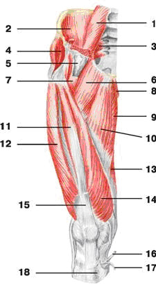
Rice. 74.
A - front view: 1 - iliopsoas muscle; 2 - pectineus muscle; 3 -
adductor longus; 4 -
thin muscle; 5 - sartorius muscle; 6 -
medial vastus muscle; 7 - quadriceps tendon; 8 -
patellar ligament; 9 -
calf muscle; 10 -
soleus muscle; 11 -
extensor longus fingers; 12 -
long peroneus muscle; 13 -
front tibialis muscle; 14 -
vastus lateralis; 15 -
rectus femoris muscle; B - rear view: 1 - gluteus maximus muscle; 2 - iliotibial tract (part of the lata fascia of the thigh); 3-
biceps femoris muscle; 4-
calf muscle; 5 -
calcaneal (Achilles) tendon; 6 -
semimembranosus muscle; 7- semitendinosus muscle
Superior and inferior gemellus muscles(m. gemellus superior et inferior) start from the ischium and ischial tuberosity; are attached to the greater trochanter. Rotate the thigh outward.
Piriformis muscle(m. piriformis) originates from the pelvic surface of the sacrum, passes through the sciatic foramen and attaches to the greater trochanter of the femur. Rotates the hip outward, with slight abduction.
External group of pelvic muscles. The muscles of this group are divided into three layers: superficial, middle and deep. The first layer contains the gluteus maximus, tensor fascia lata, the second layer contains the gluteus medius, superior and inferior gemellus, quadratus femoris, and the third contains the gluteus minimus and obturator externus muscles.
Gluteus maximus muscle(m. gluteus maximus) starts from the iliac crest, the dorsal surface of the sacrum, coccyx and the tendon part of the erector spinae muscle; attaches to the gluteal tuberosity of the femur. Extends the thigh, turns it slightly outward, abducts the thigh, fixes the pelvis and torso.
Gluteus medius muscle(m. gluteus medius) originates from the iliac bone of the fascia lata and attaches to the greater trochanter of the femur. Abducts and rotates the hip, participates in fixing the pelvis and torso in vertical position at fixed lower limb together with the gluteus minimus muscle.
Gluteus minimus(m. gluteus minimus) originates from the ilium and attaches to the greater trochanter of the femur. Abducts and rotates the thigh inward, outward; straightens the body.
Tensor fascia lata (t. tensor fasciae latae) starts from the iliac spine, goes down and passes into the iliotibial tract of the lata fascia of the thigh. By contracting, it strains the fascia and helps strengthen the knee joint in an extended position.
Quadratus femoris(m. quadratus femoris) comes from the ischial thigh and attaches to the intertrochanteric ridge. Rotates the hip outward.
Obturator externus muscle(m. obturatorius externa) starts from the outer surface of the pubic bone, the branch of the ischium and the obturator membrane; attaches to the trochanteric fossa of the femur and articular capsule. Rotates the hip outward.
The gluteus maximus muscle (m. gluteus maximus) (Fig. 128) extends the thigh, straightens the torso bent forward, stretches the lata fascia of the thigh, and fixes the pelvis and torso in a standing position. It is large, flat, rhomboid muscle, the powerful development of which is explained by human upright posture. It starts from the back of the outer (gluteal) surface of the ilium, from the lateral edge of the sacrum and coccyx. The lower bundles of the muscle are attached to the gluteal tuberosity of the femur, and the upper bundles are woven into the iliotibial tract. Between the gluteal tuberosity and the muscle there is the trochanteric bursa of the gluteus maximus muscle (bursa trochanterica m. giutei maximi).
The gluteus medius muscle (m. gluteus medius) (Fig. 128, 130) abducts the thigh. In this case, the anterior bundles rotate the thigh inward, and the posterior bundles rotate the thigh outward. When the hip is in a fixed position, the pelvis moves to the side. Also takes part in straightening the body bent forward. This is a thick muscle located under the gluteus maximus muscle and consisting of superficial and deep layers of muscle bundles.
The beams themselves are arranged fan-shaped. The origin of the muscle is on the outer surface of the iliac wing and on the lata fascia of the thigh, and the insertion point is on the greater trochanter of the femur. The trochanteric bursa of the gluteus medius muscle (bursa trochanterica m. giutei medii) is also located here. The gluteus minimus muscle (m. gluteus minimus) (Fig. 128) abducts the thigh and takes part in straightening the body. It is covered by the gluteus medius muscle, its origin point is located on the outer surface of the iliac wing between the anterior and inferior gluteal lines. The muscle is attached to the anterior edge of the greater trochanter of the femur.
| Rice. 128. Pelvic muscles (rear view): 1 - gluteus maximus muscle; |
|
 | Rice. 129. Muscles of the pelvis and thigh (front view): 1 - psoas minor muscle; |
 | Rice. 130. Muscles of the pelvis and thigh (front view): 1 - psoas major muscle; |
The muscle that stretches the fascia lata of the thigh (m. tensor fasciae latae) (Fig. 129) strains the fascia lata of the thigh and takes part in its flexion. This flat, elongated muscle is located on the anterolateral surface of the pelvis. It starts from the superior anterior iliac spine and attaches to the iliotibial tract.
The quadratus femoris muscle (m. quadratus femoris) (Fig. 128) rotates the thigh outward. It has the shape of a rectangle, partially covered by the gluteus maximus muscle. It starts from the lateral surface of the ischial tuberosity and attaches to the greater trochanter and intertrochanteric crest of the femur. The distal end of the muscle grows into the lata fascia of the thigh.
The superior gemellus muscle (m. gemellus superis) (Fig. 128), like the quadratus muscle, rotates the thigh outward. It is a muscle cord, the origin of which is located on the ischial spine, and the attachment point is in the trochanteric fossa of the femur.
The inferior gemellus muscle (m. gemellus inferior) (Fig. 128) rotates the thigh outward. The origin of the muscle is the ischial tuberosity, and the attachment point is the trochanteric fossa of the femur.
The external locking muscle (m. obturatorius externum) (Fig. 128), together with the previous muscles, rotates the thigh outward. The muscle is an irregular triangle, its origin point is located on the outer surface of the pubic and ischial bones in the area of the locking membrane, and the attachment point is the trochanteric fossa of the femur.
Pelvic muscles
Pelvic muscles divided into two groups - internal and external. To the group internal muscles include the iliopsoas, obturator internus and piriformis muscles. The group of external muscles includes the large, middle and small gluteal muscles; tensor fascia lata, quadratus femoris and obturator externus.
Internal pelvic muscle group
Iliopsoas muscle consists of two muscles - the psoas major and the iliacus, which, starting in different places (on the lumbar vertebrae and the ilium), unite into a single muscle attached to the femur. At a great distance, both parts of the muscle take part in the formation of the muscular basis of the posterior wall of the abdominal cavity.
Psoas major muscle thick, fusiform, starts from the lateral surface of the bodies and transverse processes of the 12th thoracic and all lumbar vertebrae. Located in front of the transverse processes, this muscle is tightly adjacent to the vertebral bodies. Next, the muscle goes down, crosses the border line of the pelvis in front and connects with the iliacus muscle.
Iliacus muscle massive flat, occupies the iliac fossa, adjacent on the lateral side to the psoas major muscle. It starts from the upper two-thirds of the iliac fossa, the inner lip of the iliac crest, the anterior sacroiliac and iliopsoas ligaments.
Iliopsoas muscle exits (behind the inguinal ligament) through the muscle lacuna into the thigh area and attaches to the lesser trochanter of the femur. The muscle flexes the thigh hip joint. With a fixed lower limb, it bends the lumbar part of the spine and tilts the pelvis along with the torso forward.
Psoas minor muscle inconsistent (absent in 40% of cases). It starts from the intervertebral disc and the adjacent edges of the bodies of the last thoracic and 1st lumbar vertebrae. The muscle is located on the anterior surface of the psoas major muscle, fused with the fascia covering it. The thin belly of this muscle passes into a long tendon, which is attached to the arcuate line of the ilium and to the iliopubic eminence; part of the muscle tendon bundles passes into the iliac fascia and into the iliopectineal arch. The muscle tightens the fascia iliaca, increasing support for the iliopsoas muscle.
Obturator internus muscle starts from the edges of the obturator foramen (excluding the obturator groove), the inner surface of the obturator membrane, the pelvic surface of the ilium (above the obturator foramen) and from the obturator fascia. The muscle exits the pelvic cavity through the lesser sciatic foramen, changes direction at an acute angle, throwing itself over the edge of the lesser sciatic notch (there is a synovial bursa here and is attached to the medial surface of the greater trochanter. Upon exiting the foramen, the superior and inferior gemellus muscles join the obturator internus muscle , also attached to the greater trochanter.
Superior gemellus starts from the ischium, the inferior gemellus muscle - from the ischial tuberosity. The muscle rotates the thigh outward.
Piriformis muscle starts from the pelvic surface of the sacrum (2-4 sacral vertebrae), lateral to the pelvic sacral foramina, exits the pelvic cavity through the greater sciatic foramen. Behind the femoral neck, the muscle passes into the round tendon, which is attached to the top of the greater trochanter. The muscle rotates the thigh outward with slight abduction.
Category "Kinesiology" In this article we will look at the anatomy, function and kinesiology of the lower extremity girdle (pelvis) in particular: the iliopsoas, psoas major, psoas minor, piriformis, obturator internus and externus, gemellus superior and inferior, tensor fasciae lata, quadratus muscle thighs, coccygeus muscle. Hip flexors. Biomechanical movements of the lower limb belt in space. Exercise technique.
The legs have a more massive skeleton than the arms. Their muscles have great strength, but in a place with less variety and a limited range of movements. The muscles located in the area of the lower extremities drive the movement of the leg in the hip joint, as well as the spinal column.
Rice. 1. Muscles of the anterior wall of the abdomen and pelvis
The muscles of the lower limb girdle - the pelvis - surround the hip joint. They start from the sacrum, pelvic bones and spine, and are attached to the proximal end of the femur. Topographically, they are divided into two groups: internal and external pelvic muscles.
Let's look at the pelvic muscles from an anatomical point of view.
Rice. 2. Muscles of the pelvis and thigh (front view)
Where: 1 - psoas minor muscle; 2 - iliacus muscle; 3 - psoas major muscle; 4 - piriformis muscle;
5 - iliopsoas muscle; 6 - vascular lacuna; 7 - muscle that stretches the lata fascia of the thigh;
8 - pectineus muscle; 9 - long adductor muscle; 10 - sartorius muscle; 11 - thin muscle;
2 - the longest rectus femoris muscle; 13 - adductor magnus; 14 - iliotibial tract;
15 - vastus lateralis muscle; 16 - vastus medialis;
17 - tendon of the longest rectus femoris muscle; 18 - sartorius tendon

- Greater lumbar (lat. psoas major), originating from the XII thoracic and I-II lumbar vertebrae. Both muscles join together, pass under the inguinal ligament in the muscle lacuna and attach to the lesser trochanter of the femur. (Number 3)
Function:
flexes the hip and rotates it outward.
Innervation:
lumbar plexus, LI-SII.
- Psoas minor muscle (lat. psoas minor) is unstable, has a fusiform shape and is located on the anterior surface of the psoas major muscle. Its point of origin is on the lateral surface of the bodies of the 1st lumbar and 12th thoracic vertebrae, and its attachment point is on the iliac fascia and the crest of the pubic bone. (Number 1)
Function:
stretches the fascia iliaca.
Innervation:
lumbar plexus, LI-LII.
- Piriformis muscle (lat. piriformis) rotates the thigh outward and takes part in its abduction. The muscle has the shape of a flat isosceles triangle. It starts from the anterior (pelvic) surface of the sacrum, exits the pelvic cavity through the greater sciatic foramen and is attached to the apex of the greater trochanter pelvic bone. At the site of muscle attachment there is a mucous bursa of the piriformis muscle (bursa m. piriformis). The muscle completely fills the large sciatic foramen, forming supragiriform spaces on top and infrapiriform spaces below, through which vessels and nerves pass. (Number 4)
Function:
externally rotates the hip.
Innervation:
sacral plexus,SI-SII.
Rice. 3. Pelvic muscles (back view)

- Internus obturator muscle (lat. obturatorius internus) rotates the hip outward. This is a flat muscle with fan-shaped bundles. Its starting point is located on the inner surface of the pelvic bone in the circumference of the locking membrane. The muscle exits the pelvic cavity through the lesser sciatic foramen and is attached to the vertical fossa of the femur. Between the muscle and the locking groove of the pubic bone, a small gap is formed - the locking canal (canalis obturatorius), through which the vessels and nerve pass. (Fig.3)
Function:
externally rotates the hip.
Innervation:
sacral plexus, LI-SII.
- Superior and inferior gemellus muscles (lat. gemellus superior et inferior) begin from the ischial spine (upper) and the ischial tuberosity (lower); are attached in the trochanteric fossa. (Fig.3)
Function:
rotate the hip outward.
Innervation:
sacral plexus, LIV-SII
- Coccygeus muscle (lat. coccygeus), contracting, takes part in strengthening the walls of the pelvis. The muscle is rudimentary, it is a thin plate with a small number of muscle bundles. Its point of origin is located on the ischial spine, and its attachment point is on the outer surface of the two lower sacral and two or three upper coccygeal vertebrae.
Basic hip flexors
- iliopsoas,
- rectus femoris muscle,
- sartorius muscle and
- tensor fascia lata.
Acting synergistically, these muscles cause flexion of the hip joint, such as when raising a straight leg and knee. They also contract eccentrically to control hip extension, such as during the downward phase of a straight leg or knee raise. Let's look at each one separately.
Rice. 4. Muscles of the pelvis and thigh (front view)
 Iliopsoas muscle(Fig.4) flexes the thigh at the hip joint, rotating it outward. When the hip is in a fixed position, it bends the lumbar region and pelvis, tilting the torso forward. Received its name from the site of its origin on the inner surface of the ilium; it is attached to the lesser trochanter of the femur.
The muscle is formed as a result of the connection of the psoas major muscle ( lat. psoas major) and iliacus muscle ( lat. iliacus).
The psoas major muscle is longus muscle fusiform, starting from the lateral surface of the bodies of the I-IV lumbar vertebrae and the XII thoracic vertebra. The iliacus muscle has the shape of a triangle and fills the iliac fossa, on the walls of which is the point of origin of the muscle. Both muscles connect at the attachment point, which is located on the lesser trochanter of the femur. Between the joint capsule and the muscle tendon is the iliopectineal bursa (bursa iliopectinea). Essentially, it consists of three muscles: the round major and minor (absent in about 10% of the population) psoas and iliacus, which function as a single unit.

Kinesiology: The psoas muscle must exert considerable force to raise and lower the mass of the straightened leg. For most people abdominal muscles not strong enough and cannot balance the force generated by the psoas muscle to keep the spine in a neutral position when lifting the straight leg. This is one of the reasons why it is not recommended to lift the body from a lying position without the help of arms and legs with straight legs. Because the psoas muscle originates in the lumbar spine, stiffness or hypertrophy can result in passive hyperextension of the lumbar spine.
Stiffness in the iliopsoas muscle can be attributed to insufficient stretching exercises, as well as improper posture while standing or sitting. To stretch the iliopsoas muscle, the client should stand in a lunge forward with one leg bent at the knee and the heel of the other leg without touching the floor. Then, contracting the abdominal muscles, he must bend the lumbar spine and fix this position for at least 10 seconds. You should carefully monitor how the client performs this exercise, as there is a tendency to hyperextend the lumbar spine, which is accompanied by unnecessary stress on it.
To strengthen the iliopsoas muscle, from a lying position on your back, use your abdominal muscles to lift your pelvis up to stabilize your lower back and then alternately lift one straight leg and then the other leg up.
Rice. 5. Thigh. (Front view)

- Rectus femoris muscle(Fig. 5)
Quadriceps femoris (lat. quadriceps femoris) is located on the front surface of the thigh and consists of 4 heads - muscles. Since one of the four heads is involved to a greater extent in flexing the pelvis main muscle- rectus bera muscle, let’s look at it in more detail
Rectus femoris muscle(lat. musculus rectus femoris) is the longest of all muscle heads. Occupies the anterior surface of the thigh. It begins with a thin tendon from the lower anterior spine, the supraacetabular groove. At the very beginning m is covered. tensor fasciae latae and sartorius muscle y. It goes down and passes into a narrow tendon, which is part of the common tendon of the quadriceps muscle. Having reached tibia the tendon attaches to the tibial tuberosity. Below the patella it is called the patellar ligament (lat. ligamentum patellae).
The only one of the four muscles of the quadriceps group that crosses the hip joint. Concentric contraction of this muscle results in hip flexion, knee extension, or both. The best exercise to strengthen this muscle, lift the straight leg from a standing position. To stretch the rectus femoris, perform an iliopsoas stretch and then lower your torso until the knee of your back leg is bent.
- Sartorius(Fig. 5)
Sartorius (lat. sartorius) the longest muscle in the human body, starting from the superior anterior iliac spine; attaches to the medial surface of the tibial tuberosity.
Function:
flexes the thigh and lower leg, bent in knee joint the limb rotates inward.
Innervation:
femoral nerve, LI-LII.
This multi-joint muscle flexes, abducts and externally rotates the hip joint and at the same time flexes and internally rotates the knee joint. Lateral to the sartorius muscle is the tensor fascia lata - short muscle with very long tendon, which connects to the lower fibers of the gluteus maximus muscle. The tensor fascia lata originates on the anterior superior iliac bone and inserts on the lateral aspect of the tibia below the knee.
- (Fig. 5)
Tensor fascia lata (lat. Musculus tensor fasciae latae)
A flat, slightly elongated muscle that lies on the anterolateral surface of the pelvis. With its distal end it is woven into the fascia lata of the thigh. The muscle begins on the outer lip of the iliac crest, closer to the superior anterior iliac spine. The muscle bundles are directed vertically downwards, passing into the iliotibial tract of the fascia lata of the thigh.
Functions: Stretches the fascia lata and the iliotibial band. Through it it acts on the knee joint and flexes the hip. Thanks to the connection with the tensioner fascia lata The hip muscles, gluteus maximus and gluteus medius, contribute to movement of the knee joint. This muscle is not only a hip flexor, but also a pronator. In addition, it abducts the hip. When the hip is fixed, it participates in the rotation of the pelvis.
The iliopsoas muscle (m. iliopsoas) flexes the thigh at the hip joint, rotating it outward. When the hip is in a fixed position, it bends the lumbar region and pelvis, tilting the torso forward. The muscle is formed as a result of the connection of the psoas major muscle (m. psoas major) and the iliac muscle (m. iliacus). The psoas major muscle is a long, fusiform muscle, starting from the lateral surface of the bodies of the I–IV lumbar vertebrae and the XII thoracic vertebra. The iliacus muscle has the shape of a triangle and fills the iliac fossa, on the walls of which is the point of origin of the muscle. Both muscles connect at the attachment point, which is located on the lesser trochanter of the femur. Between the joint capsule and the muscle tendon is the iliopectineal bursa (bursa iliopectinea).The psoas minor muscle (m. psoas minor) stretches the fascia iliaca. The muscle is unstable, has a fusiform shape and is located on the anterior surface of the psoas major muscle.
Its point of origin is on the lateral surface of the bodies of the 1st lumbar and 12th thoracic vertebrae, and its attachment point is on the iliac fascia and the crest of the pubic bone.
The piriformis muscle (m. piriformis) rotates the thigh outward and takes part in its abduction. The muscle has the shape of a flat isosceles triangle. It starts from the anterior (pelvic) surface of the sacrum, exits the pelvic cavity through the greater sciatic foramen and attaches to the top of the greater trochanter of the pelvic bone. At the site of muscle attachment there is a mucous bursa of the piriformis muscle (bursa m. piriformis). The muscle completely fills the large sciatic foramen, forming supragiriform spaces on top and infrapiriform spaces below, through which vessels and nerves pass.
The internal locking muscle (m. obturatorius internus) rotates the thigh outward. This is a flat muscle with fan-shaped bundles. Its starting point is located on the inner surface of the pelvic bone in the circumference of the locking membrane.
The muscle exits the pelvic cavity through the lesser sciatic foramen and is attached to the vertical fossa of the femur. Between the muscle and the locking groove of the pubic bone, a small gap is formed - the locking canal (canalis obturatorius), through which the vessels and nerve pass.
The coccygeal muscle (m. coccygeus), contracting, takes part in strengthening the walls of the pelvis. The muscle is rudimentary, it is a thin plate with a small number of muscle bundles. Its point of origin is located on the ischial spine, and its attachment point is on the outer surface of the two lower sacral and two or three upper coccygeal vertebrae.
Muscles of the pelvis and thigh (front view):
1 - psoas major muscle;
2 - iliacus muscle;
3 - piriformis muscle;
4 - gluteus medius muscle;
5 - iliopectineal bursa;
6 - pectineus muscle;
7 - iliopsoas muscle;
8 - thin muscle;
9 - adductor magnus;
10 - long adductor muscle;
11 - vastus intermedius;
12 - vastus lateralis muscle;
13 - semimembranosus muscle;
14 - vastus medialis muscle;
15 - tendon of the longest rectus femoris muscle;
16 - semitendinosus tendon;
17 - tendon of the thin muscle;
18 - tendon of the sartorius muscle.


