Muscles of the back and pelvis anatomy. Topic: “Muscles of the lower limb
The pelvic muscles, starting on the bones of the pelvis and spinal column, surround the hip joint and attach to the upper end femur. For ease of study, the pelvic muscles are divided into internal and external groups.
Inner group
1. Ileo- psoas(m. iliopsoas; see Fig. 70) consists of two separate muscles that connect only at the point of attachment: the lumbar major (m. psoas major) and the iliac muscle (m. iliacus). Origin: the psoas major muscle - from the lateral surface of the bodies of the XII thoracic and I - IV lumbar vertebrae, as well as from their transverse processes, the iliacus muscle - from the walls of the iliac fossa; insertion: lesser trochanter of the femur.
Function: flexes the thigh and rotates it outward. When the hip is fixed, it flexes lumbar region spine and pelvis.
2. The psoas minor muscle (m. psoas minor; see Fig. 70) is unstable. Beginning: lateral surfaces of the bodies of the XII thoracic and I lumbar vertebrae; attachment: fascia iliaca, pubic crest.
Function: stretches the fascia iliaca.
3. Piriformis muscle (m. piriformis; Fig. 81). Origin: pelvic surface of the sacrum: insertion: apex of the greater trochanter of the femur, after exiting the pelvic cavity through the greater sciatic foramen.
Function: rotates the thigh outward.
4. Internal obturator muscle (m. obturator internus; see Fig. 81). Beginning: the inner surface of the pelvic bone in the circumference of the obturator membrane; insertion: trochanteric fossa of the femur, after exiting the pelvis through the lesser sciatic foramen.
Outdoor group
1. The gluteus maximus muscle (m. gluteus maximus; Fig. 82) reaches greatest development in a person due to vertical position bodies. Origin: gluteal surface of the ilium, dorsal surface of the sacrum and coccyx, thoracolumbar fascia; attachment: gluteal tuberosity of the femur, part of the fibers passes into the iliotibial tract.
Function: extends the hip, when standing, fixes the pelvis, and with it the torso.

2. Gluteus medius muscle (m. gluteus medius; see Fig. 82). Origin: gluteal surface of the ilium, fascia lata of the thigh; insertion: greater trochanter of the femur.
Function: abducts the thigh, the anterior bundles rotate the thigh inward, the posterior bundles rotate the thigh outward. When fixing the hip, the pelvis tilts to the side. Participates in straightening the body bent forward.
3. Gluteus minimus (m. gluteus minimus; see Fig. 82). Origin: ilium, between the anterior and inferior gluteal lines; insertion: anterior edge of the greater trochanter of the femur.
Function: abducts the thigh, straightens the torso.
4. Tensor fasciae latae (m. tensor fasciae latae; Fig. 83). Origin: superior anterior iliac spine; insertion: iliotibial tract.

Function: annoying fascia lata hips, flexes the hip.
5. Quadratus femoris (m. quadratus femoris; see Fig. 82). Origin: ischial tuberosity; insertion: greater trochanter and intertrochanteric crest of the femur.
Function: rotates the thigh outward.
6. Superior gemellus muscle (m. gemellus superior; see Fig. 82). Origin: ischial spine; insertion: trochanteric fossa of the femur.
Function: rotates the thigh outward.
7. Lower gemellus muscle (m. gemellus inferior; see Fig. 82). Origin: ischial tuberosity; insertion: trochanteric fossa of the femur.
Function: rotates the thigh outward.
8. External obturator muscle (m. obturator externus; see Fig. 81). Beginning: the outer surfaces of the pubis and ischium around the obturator membrane; insertion: trochanteric fossa of the femur.
Function: rotates the thigh outward.
The pelvic muscles are divided into internal and external groups.
Internal pelvic muscle group
- Psoas major muscle, m.psoas major.
- Psoas minor muscle, m. psoas minor.
- Iliacus muscle, m. iliacus.
- Iliopsoas muscle, m. iliopsoas.
- Obturator internus muscle, m. obturatorius interims.
- Piriformis muscle, m. piriformis.
- Coccygeus muscle, m. coccygeus.
External pelvic muscle group
- Gluteus maximus muscle, m. gluteus maximus.
- Gluteus medius muscle, m. gluteus medius.
- Gluteus minimus, m. gluteus minimus.
- Quadratus femoris muscle, m. quadratus femoris.
- Superior gemellus muscle, m.gemellus superior.
- Gemini inferior muscle, m. gemellus inferior.
- Obturator externus muscle, m. obturatorius extemus.
- Tensor fascia lata, m. tensor fasciae latae.
Internal pelvic muscle group
- Psoas major muscle, m. psoas major, long, fusiform, begins with 5 teeth from the lateral surface of the bodies of the XII thoracic, four upper lumbar vertebrae and the corresponding intervertebral cartilages. The deeper muscle bundles originate from the transverse processes of all lumbar vertebrae. Tapering somewhat, the muscle is directed downward and slightly outward and, connecting with the bundles of the iliacus muscle, m. iliacus, forms the common iliopsoas muscle, m. iliopsoas.
- Psoas minor muscle, m. psoas minor (fickle), thin, spindle-shaped, located on the anterior surface of the m. psoas major. It starts from the lateral surface of the bodies of the XII thoracic and I lumbar vertebrae and, going down, passes with its tendon into the fascia iliaca, attaching with it to the pecten ossis pubis and to the eminentia iliopectinea. Action: stretches the fascia iliaca. Innervation: rr. musculares plexus lumbalis (L1-L2). Blood supply: aa. lumbales.
- Iliacus muscle m. iliacus, fills the entire iliac fossa, fossa iliaca, originating from its walls. The shape of the muscle approaches a triangle, with its apex facing downwards. The bundles that make up the muscle fan-like converge to the linea tenninalis and here merge with the bundles of m. psoas major, forming m. iliopsoas.
- Iliopsoas muscle, m. iliopsoas, is formed as a result of the connection of the distal muscle bundles of m. iliacus and m. psoas major. The muscle leaves the pelvic cavity through the lacuna musculorum and, moving downwards, passes along the anterior surface hip joint, attached by a thin short tendon to the trochanter minor femoris; between the joint capsule and the muscle tendon there is an iliopectineal bursa, bursa ileopectinea, which often communicates with the cavity of the hip joint. Action: flexes the thigh at the hip joint, rotating it outward. When the hip is fixed, it tilts (bends) the torso forward. Innervation: rr. musculares plexus lumbalis (L1-L2). Blood supply: aa. iliolumbalis, circumflexa ilium profunda.
- Obturator internus muscle, m. obturatorius internus, is a flattened muscle in which the muscle bundles are directed slightly fan-shaped. The widest part of the muscle originates from inner surface pelvic bone in the circumference of the membrana obturatoria and from its inner surface. A small gap between the muscle bundles and the sulcus obturatorius of the pubic bone turns into the obturator canal, canalis obturatorius. through which blood vessels and nerves pass. Then the muscle bundles, converging, are directed outward and, bending almost at a right angle through the lesser sciatic notch, leave the pelvic cavity through the foramen ischiadicum minus, attaching with a short, powerful tendon in the fossa trochanterica area. At the point of inflection over the edge of the lesser sciatic notch there is an ischial bursa of the obturator internus muscle, bursa ischiadica m. obluratorii interni. Topographically, the obturator internus muscle is divided into two parts: the large one, before exiting the pelvic cavity, the intrapelvic muscle, and the smaller, tendinous muscle, which lies under the greater muscle. gluteal muscle, - extrapelvic. Action: supinates the thigh. Innervation: rr. musculares plexus sacralis. Blood supply: aa.glutea inferior, obturatoria, pudenda intema.
- Piriformis muscle, m. piriformis, has the appearance of a flat isosceles triangle, the base of which originates from the anterior surface of the sacrum, lateral to the openings between II and IV foramina sacralia pelvina. Converging, the muscle bundles are directed outward, exit the pelvic cavity through the greater sciatic foramen, foramen ischiadicum majus, and, passing into a narrow and short tendon, are attached to the top of the trochanter major. At the site of muscle attachment there is a mucous bursa of the piriformis muscle, bursa musculi piriformis. Passing through the greater sciatic foramen, the muscle does not completely fill it, leaving small gaps along the upper and lower edges through which blood vessels and nerves pass. The gap located along the upper edge of the piriformis muscle is called the suppiriformis foramen, and along the lower edge - the infrapiriformis foramen. Action: supinates the thigh and also participates in its abduction. Innervation: rr. musculares plexus sacralis.Blood supply: aa. gluteae, superior et inferior.
- The coccygeal muscle, m.coccygeus, is a thin plate containing relatively few muscle bundles. Originating from spina ischiadica, the muscle follows inside lig. sacrospinale and is attached to the outer surface of the 2 lower sacral and 2-3 upper coccygeal vertebrae. Action: in humans this muscle is rudimentary; during contraction, it takes part in strengthening the walls of the pelvis. Innervation: rr. musculares nervi pudendi.Blood supply: rr. musculares a. pudendae intemae.
External pelvic muscle group
- Gluteus maximus muscle, m. gluteus maximus, approximates a rhombus in shape. The muscle is large-fiber, powerful, flat and reaches a thickness of 2-3 cm. In its course, the muscle overlaps the greater trochanter, as well as the rest of the muscles of this group. It starts from the posterior part of the outer surface of the ilium, posterior to the linea glutea posterior, from the lateral edge of the sacrum and coccyx and from the lig. sacrotu-berale. The muscle bundles stretch obliquely downwards and laterally and are attached with their upper bundles to the fascia lata, which passes into the tractus iliotibialis, and with their lower bundles to the tuberositas glutea femoris; here between the greater trochanter and the muscle there is a trochanteric bursa of the gluteus maximus muscle, bursa trochanterica m.glutei maximi. Action: straightens the torso bent forward, straightens the hip, and also stretches the lata fascia of the thigh. Innervation: n. gluteus inferior (plexus sacralis) (L5 S1-S2). Blood supply: aa. gluteae superior et inferior, circumflexa femoris medialis, profunda femoris (perforans I).
- Gluteus medius muscle, m. gluteus medius, located under the gluteus maximus muscle. The shape is close to a triangle. The muscle is thick, and there are two layers of bundles: superficial and deep. The muscle bundles are arranged in a fan-shaped manner, starting with a wide part from the outer surface of the ilium wing, limited in front by the linea glutea anterior, above by the crista iliaca and below by the linea glutea posterior. Then all muscle bundles converge into a common powerful tendon, attached to the top of the trochanter major, where there is a trochanteric bursa of the gluteus medius muscle, bursa trochanterica m. glutei medii.Action: abducts the thigh, with the anterior bundles rotating the thigh inward, and the posterior bundles rotating outward; takes part in straightening the body bent forward. Innervation: n. gluteus superior (plexus sacralis) (L1-L5, S1). Blood supply: aa. glutea superior et circumflexa femoris lateralis.
- Gluteus minimus, m. gluteus minimus, resembles the previous one in shape, but is much thinner in diameter. Throughout its entire length, the muscle is covered by the gluteus medius muscle. The muscle begins from the outer surface of the wing of the ilium, between the linea glutea anterior and linea glutea inferior. Then the muscle bundles, converging, pass into a tendon that attaches to the anterior edge of the trochanter major; here there is the trochanteric bursa of the gluteus minor muscle, bursa trochanterica m. glutei minimi. Action: similar to the action of the gluteus medius muscle: abducts the leg and takes part in straightening the bent torso. Innervation: n. gluteus superior (plexus sacralis) (L1-L5 S1). Blood supply: aa. glutea superior, circumflexa femoris lateralis.
- Quadratus femoris muscle, m. quadratus femoris, has the appearance of a relatively thick rectangle, covered at the back by m. gluteus maximus. The muscle begins from the lateral surface of the tuber ischiadicum and attaches to the crista intertrochanterica, reaching the trochanter major. Action: rotates the thigh outward. Innervation: n. ischiadicus (plexus sacralis) (L4-L5, S1).Blood supply: aa. glutea inferior, circumflexa femoris medialis, obturatoria.
- Gemini superior muscle, m. gemellus superior, has the appearance of a small muscle cord originating from the spina ischiadica and attached to the fossa trochanterica. The muscle is adjacent to the upper edge of the tendon m. obturatorius intemus after its exit from the pelvic cavity. Action: rotates the hip outward. Innervation: branches of the plexus sacralis (L4-L5, S1). Blood supply: aa. glutea inferior, pudenda interna.
- Gemini inferior muscle, m. gemellus inferior, similar in shape to the previous one and located below the tendon of m. obturatorius intemus. The muscle starts from the tuber ischiadicum, attaches to the fossa trochanterica. Action: rotates the thigh outward. The innervation and blood supply are the same as the superior gemellus muscle.
- Obturator externus muscle, m. obturatorius externus, has the shape of an irregular triangle. It starts from the membrana obturatoria and the bony edge of the foramen obturatum with its wider part, then the muscle bundles, fan-shaped, pass into the tendon adjacent to the posterior surface of the capsule of the hip joint. The muscle is attached to the fossa trochanterica, next to the internal muscle of the same name. Action: rotates the thigh outward. Innervation: n. obturatorius (plexus lumbalis) [(LJ, 1-3-5)].Blood supply: aa. obturatoria, circumflexa femoris lateralis.
- Tensor fascia lata, m. tensor fasciae latae, flat, slightly elongated; lies on the anterolateral surface of the pelvis; its distal end is woven into the fascia lata of the thigh. The muscle begins on the labium externum cristae iliacae, closer to the spina iliaca anterior superior. The muscle bundles are directed vertically downwards, passing into the tractus iliotibialis fasciae latae. Action: strains the fascia lata of the thigh, and also takes part in hip flexion. Innervation: n. gluteus superior (plexus sacralis) (L4-L5, S1). Blood supply: aa. glutea superior, circumflexa femoris lateralis.


The pelvic area includes the pelvic bones, sacrum, coccyx, as well as ligaments, joints and membranes. Some experts also include the buttock area.
The article discusses the anatomy of the pelvis: muscles, genital and excretory organs.
Pelvic skeletal system
The pelvic skeleton consists of the pelvic bones, the sacrum and the coccygeal bone. Each of them is firmly fixed. The ilium, as well as the coccygeal bone, articulates with the sacrum.
The pelvis is divided into large and small sections.
The first consists of the sides with the wings of the ilium. On the inner surface there is the iliac fossa, and on the outside there are the gluteal fossa.
The small pelvis consists of a cylindrical cavity with upper and lower openings (that is, inlet and outlet).
The coccygeal bone is slightly mobile, which helps women during childbirth. The bony anatomy of the pelvis has the following differences in men and women:
- the male pelvis is long and narrow, in women it is shorter and wider;
- the male pelvic cavity has a conical shape, the female pelvic cavity is cylindrical;
- The wings of the ilium are more vertical in men, and more horizontal in women;
- the branches of the pubic bones in men make an angle of 70-75 degrees, in women - 90-100 degrees;
- in men the shape of the entrance resembles a heart (as on cards), in women it is round, although it happens that in women there is an entrance like a “card heart”.

Ligaments
Well-developed ligaments fix four of which are discussed above. Three joints help connect them to each other: the pubic fusion (two unpaired), the sacroiliac (pair) and the sacrococcygeal fusion.
One is located on the top edge, the other on the bottom. The third ligaments strengthen the joints of the sacrum and ilium.
Muscular system of the pelvis
In this section, the anatomy of the pelvis is presented by the parietal and visceral muscles. In the first part, in the large pelvis there is a muscle consisting of three interconnected m.iliacus, m.psoas major and m.psoas minor. In the small pelvis, the same parietal muscles are represented by the piriformis, obturator internus and coccygeus muscles.
Visceral muscles take part in the formation. It includes paired and unpaired m.sphincter ani extremus.
Here are the pubococcygeus muscle, the iliococcygeus muscle, as well as the powerfully developed orbicularis muscle of the distal part of the rectum.

Blood supply and lymphatic system
Blood enters the pelvis (anatomy here involves the participation of the pelvic walls and internal organs) from the hypogastric artery. It is divided first into anterior and posterior, and then into other branches.
IN soft fabrics of the pelvis, blood enters through a single vessel a.iliolumbalis, which branches into two terminal branches.
The walls of the pelvis are supplied by four arteries:
- lateral sacral;
- obturator;
- superior gluteal;
- inferior gluteal.
The vessels of the abdominal walls and retroperitoneal space are involved in the roundabout circulation. In the circuitous venous circle, the main veins pass between the large and small pelvis. There are abundant venous anastomoses located next to the wall of the rectum and in its thickness, as well as under the peritoneum of the pelvis. When large pelvic veins are blocked, the veins of the spine, lumbar, anterior abdominal wall and retroperitoneal tissue.
The anatomy of the pelvis, like other systems, suggests variability in the morphology of blood vessels in the lymph.
The main lymphatic collectors from the pelvic organs are the iliac lymphatic plexuses, which divert lymph.
Lymphatic vessels under the peritoneum mainly pass at the level of the middle floor of the pelvis.
Innervation
The nerves in this area are divided into:
- somatic;
- autonomic (parasympathetic and sympathetic).
The somatic nerve system is represented by those connected to the lumbar. Sympathetic - the sacral part of the border trunks and the unpaired coccygeal ganglion. Parasympathetic nerves are nn.pelvici s.splanchnici sacrales.

Buttocks
The anatomy of the gluteal region is often not included in the pelvis. However, topographically it should be classified here and not to the lower extremities. Therefore, we will briefly touch on this too.
Above gluteal region bounded by the iliac crest, and below by the gluteal fold, under which is the gluteal groove. On the lateral side, you can imagine a vertical line of a row of bones, and on the medial side, both areas are separated by the intergluteal gap.
Let's look at the anatomy here layer by layer:
- the skin of this area is thick and dense;
- well-developed subcutaneous tissue with superficial, middle and lower nerves;
- then follows the superficial plate of the gluteal fascia;
- gluteus maximus muscle;
- plate of the gluteal fascia;
- fatty tissue between big muscle and the middle layer of muscles;
- middle layer of muscles;
- deep layer of muscles;
- bones.

Excretory organs
The anatomy of the pelvis includes an unpaired muscular organ - the bladder. It consists of an apex, body, bottom and neck. One department here merges into another. The fundus is fixed with a urogenital diaphragm. When the bladder begins to fill, its shape becomes ovoid. When the bladder is empty, the shape is close to saucer-shaped.
The blood supply comes from the hypogastric artery system, and the venous outflow is directed into the dense cystic plexus, which is adjacent to the lateral surfaces and the prostate gland.
Innervation is carried out by somatic and autonomic fibers.
The rectum begins to develop from embryonic rudiments. The upper section is derived from the endoderm, and the lower section appears by incising from the surface of the ectodermal layer.
The rectum is located at the level of the posterior pelvis. It is divided into three sections: upper, middle and lower.
The muscles on the outside are represented by powerful longitudinal fibers, and on the inside by circular fibers. The mucous membrane consists of numerous folds. The innervation here is similar to that in the bladder.
Reproductive system
Without the reproductive system, it is impossible to consider the pelvis (structure). The anatomy of this region in both sexes consists of the gonad, Wolffian body, canal, Müllerian duct, urogenital sinus and genital tubercles, folds and ridges.
The sex gland is laid in the lower back and turns into a testicle or ovary, respectively. The Wolffian body, canal and Müllerian duct are also formed here. However, the Müllerian canals are further differentiated in the female sex, and the Wolffian body and ducts in the male sex.
The remaining rudiments are reflected on the external organs.
The testicle and ovary grow behind the peritoneum.

The male reproductive system is represented by:
- testicular integument, consisting of skin, tunica dartos, cooper's fascia, cremaster, common and proper tunica vaginalis, tunica albuginea;
- seminal gland;
- lymphatic system;
- an appendage consisting of three sections (head, body and tail);
- spermatic cord;
- seminal vesicles (hollow tubes with bay-shaped protrusions);
- (glandular-muscular organ between the diaphragm and the bottom of the bladder);
- the penis, consisting of three sections (root, body and head);
- urethra.
The pelvis includes the reproductive system of:
- uterus (derivative of Müllerian canals);
- ovaries located in a special ovarian fossa;
- fallopian tubes, consisting of four sections (funnel, dilated part, isthmus and part piercing the wall);
- vagina;
- external genitalia, consisting of the labia majora and vulva.

Crotch
This area is located from the pubic hill to the top of the coccygeal bone of the pelvis.
The anatomy of the perineum in both men and women is divided into 2 areas: the pudendal (anterior) and the anal (posterior). In front of the region corresponds to the genitourinary triangle, and behind it corresponds to the rectal triangle.
Conclusion
This is the structure that represents the pelvis as a whole. The anatomy of this area is certainly the most complex system. The article gives only short review what it consists of and how it works.
Gas muscles include those muscles that begin at pelvic bones and are attached to the femur, i.e., they act on the hip joints.
Anterior pelvic muscles
The iliac muscle (m. iliacus) (Fig. 196) begins on the entire area of the fossa iliaca of the ilium, then under the inguinal ligament in the lacuna musculorum in the place with m. psoas major passes in front of the femoral head. Attaches to the lesser trochanter of the femur. Between the joint capsule and the deep layer of muscle fascia there is a mucous bursa, which in some cases communicates with the joint cavity.
196. Muscles of the pelvis and thigh, front.
1 - m. psoas major; 2 - m. piriformis; 3 - lig. inguinal; 4 - lacuna vasorum; 5 - m. pectineus; 6 - m. adductor longus; 7 - m. gracilis; 8 - m. vastus medialis; 9 - lig. patellae; 10 - tendo m. recti femoris; 11 - tractus iliotibialis; 12 - m. vastus lateralis; 13 - m. tensor fasciae latae; 14 - m. sartorius; 15 - m. iliacus.
Innervation: branches of plexus lumbalis and n. femoralis LII-IV.
Function. When free lower limb the muscle flexes the thigh at the hip joint. When standing, the torso bends forward. Functionally combined with m. psoas major.
Posterior pelvic muscles
The gluteus maximus muscle (m. gluteus maximus) is well developed, massive, irregularly quadrangular in shape. Starts from the posterior 2/3 of the iliac crest, f. lumbodorsalis, lateral parts of the sacrum, coccyx and pelvic ligaments. Large muscle bundles separated by layers connective tissue, are directed laterally and downwards. The anterior muscle bundles form a flat tendon covering the front of the greater trochanter of the femur, and then pass into the tendon tract of the thigh. The posterior bundles are attached to the tuberositas glutea of the femur. Between the tendon and the greater trochanter there is a mucous bursa.
Innervation: n. gluteus inferior (LV-SI).
Function. With a free lower limb, the thigh extends at the hip joint. When standing, the torso extends.
Gluteus medius muscle (m. gluteus medius) (); its posterior part is covered by the previous muscle, and its anterior part is covered with thick fascia. It starts from the ilium between the linea glutea anterior et posterior below, the iliac crest behind and above. Its anterior bundles begin from the fascia lata. Attached by a broad tendon to the apex and outer surface of the greater trochanter. At the site of muscle attachment there is a mucous bursa.
Innervation: n. gluteus superior (LIV-SI).
Function. When the muscle contracts, the free part of the lower limb is abducted; when the anterior bundles contract, the thigh rotates inward (pronation), and the posterior bundles rotate outward (supination). When the lower limbs are fixed (standing), the torso tilts in the direction of muscle contraction.
The gluteus minimus muscle (m. gluteus minimus) is covered by the previous muscle, somewhat smaller in size. It begins in a fan-shaped manner from the outer surface of the wing of the ilium between the anterior and lower gluteal lines. Attached to the anterior part of the greater trochanter, i.e., in front of the attachment of m. gluteus medius, where there is a mucous bursa.
Innervation: n. gluteus superior (LIV-V-SI).
Piriformis muscle (m. piriformis); its initial part is located in the pelvic cavity. Starts from the anterior surface of the II - IV sacral vertebrae near the anterior sacral foramina, heading laterally through the greater sciatic foramen, leaves the pelvic cavity and exits onto it back surface. Attaches to the anterior edge of the greater trochanter of the femur. Above and below the piriformis muscle in the sciatic foramen there are forr. supra- et infrapiriformis.
Innervation: n. gluteus superior (SI-II).
Function. Abducts the hip with the free leg. When standing, tilts the torso in the direction of contraction.
The internal obturator muscle (m. obturatorius internus) begins on the inner surface of the small pelvis in the area of the obturator membrane and the bones surrounding it. The muscle is directed backward and, bending around the sciatic notch in the area of the lesser sciatic foramen, exits at a right angle to the posterior surface of the pelvis below m. piriformis. Attached to the fossa of the greater trochanter of the femur.
Innervation: r. muscularis plexus sacralis (LIV-SII).
Function. Abducts the hip and supinates with the lower limb free.
The superior and inferior twin muscles (mm. gemelli superior et inferior) begin from the spina ischiadica and tuber ischii, respectively, and are attached to the fossa of the greater trochanter. Both muscles are fused with the obturator internus muscle.
Innervation: rr. musculares plexus sacralis (LIV-SI).
Function. Participates in hip abduction.
Quadratus femoris muscle (m. quadratus femoris); its shape fully corresponds to its name. It starts from the ischial tuberosity and is located in the horizontal plane. Attached to the posterior part of the greater trochanter and to the crista intertrochanterica.
Innervation: rr. musculares plexus sacralis (LIV-V-SI).
Function. Due to the significant torque, it develops great force. With a free leg, the hip supinates. When standing, the pelvis tilts back.
Lateral pelvic muscles
The tensor fasciae latae is located behind the beginning of the sartorius muscle of the thigh. Its thin muscle belly is enclosed in a dense fascia of the thigh. It starts from the iliac crest behind the spina iliaca anterior superior and, going down to the thigh, continues into the tendon aponeurosis in the form of tr. iliotibialis f. latae, which ends on the lateral condyle of the tibia.
Innervation: n. gluteus superior (LIV-V).
Function. At free limb promotes hip flexion at the hip joint. When standing, it tightens the fascia of the thigh.
Medial muscles pelvis
The pectineus muscle (m. pectineus) starts from upper branch and the crest of the pubic bone, is attached to the initial part of the linea aspera femoris (Fig. 196). Its lateral edge borders m. iliacus and m. psoas major, forming with them fossa iliopectinea.
Innervation: n. obturatorius, n. femoralis (LII-III).
Function. Since the frontal axis of the hip joint is behind the point of passage of the muscle, it can not only adduct the hip, but also internally rotate and flex the hip joint. When standing, tilts the pelvis forward.
The external obturator muscle (m. obturatorius externus) is located under the m. pectineus. It can also be seen after removing m. quadratus femoris. It starts from the membrana obturatoria and the bones that form the obturator foramen. It is directed laterally and upward, bending around the back of the femoral neck. Attached to the fossa of the greater trochanter.
Innervation: n. obturatorius (LIII-IV).
Function. The free lower limb is rotated outward.
The long adductor muscle (m. adductor longus) is located below the m. pectineus, separated by an intermuscular layer of connective tissue. It starts from the pubic bone between the symphysis and the pubic tubercle. Attaches to the medial lip of the middle linea aspera femoris.
Function. Adducts the femur at the hip joint.
The short adductor muscle (m. adductor brevis) is covered in front by the m. pectineus and m. adductor longus, behind - m. adductor magnus. It starts from the lower branch of the pubic bone, then diverges downwards and laterally, attaching to the upper third of the medial lip linea aspera femoris.
Between m. pectineus and m. The adductor brevis on the thigh opens to the canalis obturatorius.
Innervation: n. obturatorius (LII-III).
Function. The same as the previous muscle.
The adductor magnus (m. adductor magnus) (Fig. 195) lives up to its name. It is covered by all the previous muscles and upon examination only its medial edge is visible. It starts from the ischial tuberosity, a branch of the ischium, then attaches along the entire length of the medial lip linea aspera femoris and to the medial epicondyle of the femur. The part of the tendon that attaches to the medial epicondyle is thickened and can be easily felt on the thigh. Lateral to this tendon, i.e. closer to the midline of the thigh, at the level of its lower third there is a hole - hiatus tendineus, the edges of which contain tendon fibers that prevent the hole from changing its shape during muscle contraction. The femoral artery and vein pass through the hole into the popliteal fossa.
Innervation: n. obturatorius and branches n. ischiadicus (LII-V).
Function. Adducts the hip. Since the origin of the muscle is behind the frontal axis of the hip joint, extension is possible.
The gluteus maximus muscle (m. gluteus maximus) (Fig. 128) extends the thigh, straightens the torso bent forward, stretches the lata fascia of the thigh, and fixes the pelvis and torso in a standing position. It is large, flat, rhomboid muscle, the powerful development of which is explained by human upright posture. It starts from the back of the outer (gluteal) surface of the ilium, from the lateral edge of the sacrum and coccyx. The lower bundles of the muscle are attached to the gluteal tuberosity of the femur, and the upper bundles are woven into the iliotibial tract. Between the gluteal tuberosity and the muscle there is the trochanteric bursa of the gluteus maximus muscle (bursa trochanterica m. giutei maximi).
The gluteus medius muscle (m. gluteus medius) (Fig. 128, 130) abducts the thigh. In this case, the anterior bundles rotate the thigh inward, and the posterior bundles rotate the thigh outward. When the hip is in a fixed position, the pelvis moves to the side. Also takes part in straightening the body bent forward. This is a thick muscle located under the gluteus maximus muscle and consists of superficial and deep layers of muscle bundles.
The beams themselves are arranged fan-shaped. The origin of the muscle is on the outer surface of the iliac wing and on the fascia lata of the thigh, and the insertion point is on the greater trochanter of the femur. The trochanteric bursa of the gluteus medius muscle (bursa trochanterica m. giutei medii) is also located here. The gluteus minimus muscle (m. gluteus minimus) (Fig. 128) abducts the thigh and takes part in straightening the body. It is covered by the gluteus medius muscle, its origin point is located on the outer surface of the iliac wing between the anterior and inferior gluteal lines. The muscle is attached to the anterior edge of the greater trochanter of the femur.
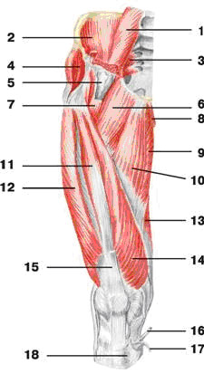
| Rice. 128. Pelvic muscles (rear view): 1 - gluteus maximus muscle; |
|
 | Rice. 129. Muscles of the pelvis and thigh (front view): 1 - psoas minor muscle; |
 | Rice. 130. Muscles of the pelvis and thigh (front view): 1 - psoas major muscle; |
The muscle that stretches the fascia lata of the thigh (m. tensor fasciae latae) (Fig. 129) strains the fascia lata of the thigh and takes part in its flexion. This flat, elongated muscle is located on the anterolateral surface of the pelvis. It starts from the superior anterior iliac spine and attaches to the iliotibial tract.
The quadratus femoris muscle (m. quadratus femoris) (Fig. 128) rotates the thigh outward. It has the shape of a rectangle, partially covered by the gluteus maximus muscle. It starts from the lateral surface of the ischial tuberosity and attaches to the greater trochanter and intertrochanteric crest of the femur. The distal end of the muscle grows into the lata fascia of the thigh.
The superior gemellus muscle (m. gemellus superis) (Fig. 128), like the quadratus muscle, rotates the thigh outward. It is a muscle cord, the origin of which is located on the ischial spine, and the attachment point is in the trochanteric fossa of the femur.
The inferior gemellus muscle (m. gemellus inferior) (Fig. 128) rotates the thigh outward. The origin of the muscle is the ischial tuberosity, and the attachment point is the trochanteric fossa of the femur.
The external locking muscle (m. obturatorius externum) (Fig. 128), together with the previous muscles, rotates the thigh outward. The muscle is an irregular triangle, its origin point is located on the outer surface of the pubic and ischial bones in the area of the locking membrane, and the attachment point is the trochanteric fossa of the femur.


