Extensor tendon of the 1st toe. Extensor hallucis brevis
Despite the apparent simplicity of the movement, flexion and extension of the toes requires the efforts of several muscles. Their coordinated work allows not only to move the toes, but also to lift the foot itself. Interestingly, there is a separate muscle for the thumb, while all the others move synchronously.
Which muscle extends the fingers
Extensor toe longus
The extensor digitorum longus is part of the anterior muscle group of the lower leg and is located along its lateral edge. This anatomical formation begins at the level of the upper third of the tibia, attaching to the anterior edge of the fibula.
Descending towards the foot, the muscle part transforms into a thin but strong tendon.
It is this that is subsequently divided into four thinner tendon bundles, one for each finger.
The place of attachment is the proximal (“near”) phalanx. At the point of fixation, it is additionally divided into three small beams, with the help of which you can use any, even the smallest part of the foot.
The long extensor also has an additional purpose. Together with the synergist muscle (third peroneus), it raises the outer edge of the foot, an action called pronation. If it is firmly fixed, the lower leg is brought closer to it.
Which muscle is responsible for flexing the fingers?

Foot muscles: dorsal and plantar
The flexor digitorum longus allows you to properly push off the surface when walking or running. This muscle formation is also involved in maintaining the balance of the entire body in a standing position.
This muscle belongs to posterior group shin and starts from back surface tibia. Descending to the foot through the heel bone, it is attached from below to the distal phalanges of the 2, 3, 4 and 5 fingers.
But do not think that the function of this muscle is limited only to the movement of the fingers.
It plays a huge role in the flexion of the foot itself and its supination.
When standing on your toes and maintaining balance in this position, you cannot do without it!
Correct formation muscle fibers, their normal tone allows the arch of the foot to occupy the correct anatomical position. This prevents the formation of flat feet.
How to check the functionality of the flexor

Functional muscle tests
At various diseases the force of muscle contraction may weaken. To find out, there are a number of simple tests:
- With one hand, the trainer or doctor holds the metatarsus in one position, and with the other, gently but firmly tries to bend the toes. The patient should try to straighten them, tensing the corresponding muscles as much as possible. In case of success muscle strength is estimated at 4 or 5 points (the maximum possible score).
- The patient lies on his back, with a soft cushion placed under his knee (you can roll up a towel). When forcibly holding the metatarsus, you must try to straighten your fingers. (2 or 3 points).
- The situation is no different from the previous one. The trainer or doctor palpates the extensor tendons. A successful attempt to straighten the fingers is awarded 1 point.
Normally, the muscle strength of a healthy person is estimated at 5 points. With insufficient nutrition or innervation of tissues, it gradually decreases.
All exercises must be done carefully to avoid spraining your ankle.
How does the thumb move?

Extensor digitorum longus stretch
The mechanics of its movements depend on several muscles at once. This complex anatomy is explained by the fact that it is this finger that largely ensures a person’s balance and makes him walk upright.
The flexor pollicis longus belongs to the posterior group of muscles of the leg and, descending to the plantar part, is transformed into a tendon.
Interestingly, in the groove between the calcaneus and talus, the flexor pollicis tendon “gives” some fibers to the flexor longus tendon.
Thus, this muscle is involved in the flexion of all toes to a greater or lesser extent.
The extensor pollicis longus belongs to the opposite, anterior group of muscles of the lower leg. At the same time, going down, it passes into the thin but very strong tendon of the long extensor.
Why muscle function may be impaired
Various diseases of muscle tissue and tendons can affect the quality of their work. Decline muscle tone can happen for several reasons:
- senile (age-related) atrophy due to metabolic disorders;
- failure of the endocrine system;
- chronic and systemic connective tissue diseases;
- fertentopathy, genetically determined diseases;
- polyneuritis, polyneuropathy of peripheral nerves;
- post-traumatic complications;
We should also talk about tendonitis. With this disease, the tissue of the tendons of the long extensor of the big toe suffers, the cause is acute inflammation. This pathological process may also involve muscle structures of various sizes. Dystrophic processes under certain favorable conditions can become chronic.
In diabetes mellitus, polyneuropathy of the lower extremities can lead to significant and serious disruption of tissue nutrition at the local level.
A frequent complication of this disease is progressive lameness, problems with trophism, up to the formation of dry gangrene.
With long-term systemic use of certain medications, persistent pain in the foot may occur. This is due to the deposition of salts in bone tissue, the formation of tumors or growths.
Hallux valgus or varus deformity of the foot is one of the main non-surgical problems in traumatology. Its curvature with deviation inward or outward not only does not look aesthetically pleasing, but also significantly reduces the quality of life.
The flexor pollicis longus muscle is located deep under the gastrocnemius and soleus muscles and
covers the tibialis posterior muscle. The muscle is located laterally on the posterior surface
shins. The muscle belly is attached to the posterior surface fibula, joining the tibialis posterior and flexor digitorum longus muscles just behind the medial malleolus.
These muscles pass through the tarsal canal formed by the medial surface of the calcaneus and the fibrous plate of the flexor tendon retinaculum. In structure and functions, this structure is identical to the carpal tunnel. The tibial artery and tibial nerve also pass through the tarsal canal.
The flexor pollicis longus, flexor digitorum longus, and tibialis posterior muscles produce inversion of the foot and plantarflexion of the ankle. In addition, the flexor pollicis longus flexes the big toe at the metatarsophalangeal and interphalangeal joints. This movement is necessary during the push-off phase of walking for efficient energy transfer. The center of gravity shifts from the heel through the foot to thumb At the end of the stance phase, the energy generated by the hips, knees and shins passes through the foot and big toe, moving the human body forward.
The flexor hallucis longus plays a significant role in channeling this energy. In addition, the flexor hallucis longus is involved in providing dynamic stabilization of the medial arch of the foot. Together with other muscles passing through the tarsal canal and the muscles of the foot, the flexor hallucis longus controls the pronation of the foot during walking, running and jumping. The flexor hallucis longus provides effective traction of the foot and helps us maintain balance effectively when walking.
Problems with this muscle can lead to flattening of the medial arch of the foot and destabilization of the ankle, causing it to appear when walking. painful sensations, loss of sensation and disturbances in walking patterns.
Palpation of the flexor hallucis longus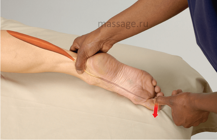
The client lies on his stomach
1. Standing at the client's feet, use your thumb to locate the medial malleolus.
2. Using a smooth sliding motion, move your thumb into the space between your ankle and Achilles tendon. (Caution: The tibial artery and nerve also pass through this area. Reposition your finger if the client experiences tingling or numbness or if you feel a pulse).
3. Three tendons are located in this area. Palpate the tendon located most
- This is the tendon of the long flexor of the big toe.
4. Ask the client to bend their thumb to make sure you have done everything correctly.
EXERCISE FOR CLIENTS: SETTING TOE STRETCH
1. Sit on the floor with your legs straight in front of you.
2. With your knees relaxed, bend at the waist and lean forward.
3. Grab your big toe with your fingers.
4. Gently pull your thumb back toward your knee for 5-10 seconds, then release.
This exercise can be done with all toes.
The muscles of the thumb recover from injury quite quickly - within one to two weeks. However, with sufficiently serious and/or chronic injuries of the thumb, there is often damage to the structure of its tendons. Tendons are practically avascular structures; their blood supply is minimal. They recover from damage approximately six times slower than muscles. In addition, it is difficult to limit the mobility of the thumb during daily activities to allow the damaged tendons to fully recover and prevent re-injury.
That is why in the process of recovery from such injuries it is extremely important the right approach to therapy. The sooner treatment begins, the faster recovery occurs. My first recommendation is not to start therapy immediately after an injury. You should wait at least 3-4 days to allow the scar tissue to fully form, and then you can use techniques that include friction (friction massage). During the first few days after injury, you can resort to light and shallow massage - this will help relieve pain and inflammation.
APPLICATION OF FRICTIONS
Based on my experience, I can confidently say that techniques that include friction are most effective in treating thumb tendon injuries. To apply friction, you can use your thumb or middle and index fingers. It is best to change fingers frequently to avoid injury yourself.
Remember that pressure should only be applied in one direction. After you have worked the tendon with friction in one direction, change the direction of the friction to the opposite direction. This will help you avoid fatigue, ensure that all tendon fibers are treated evenly, and reduce the likelihood of client discomfort.
Work the tendon fibers in one direction for 4-5 minutes, rest a little and start working in the other direction - in total you should spend about 8-10 minutes on this.
As the client's condition improves, the duration of therapy can be reduced. After applying friction massage, you can gently stretch your thumb, hand and forearm.
DETERMINATION OF THE DAMAGED AREA AND THERAPY OF INJURIES OF THE TENDONS OF THE EXTENSOR THUMB LONG AND SHORT
Determining the damaged area of the extensor pollicis longus or brevis tendon is a very simple matter. Ask the client to extend the thumb so that the entire structure is under tension. Then apply low or low friction strokes medium intensity across the fibers of the tendon of the short or long extensor pollicis (depending on the nature and location of the injury). Do this at different points along the tendon to pinpoint the location of the damaged fibers. Since the pain from these injuries does not radiate to nearby structures, the location of the damaged fibers is easy to determine by the localization of pain.
Once you have determined which part of the tendon is damaged, ask the client to relax the finger and then begin friction massage. Each stroke should completely cross the damaged area.
This approach is applicable to the treatment of any tendon injuries. Remember that the location of the damaged area should be determined quickly enough - with tendon injuries, prolonged tension 
contraindicated.
IDENTIFICATION OF THE DAMAGED AREA AND THERAPY OF INJURIES OF THE TENDONS OF THE FLEXOR THUMB BRESS AND LONGUS TENDONS
The flexor pollicis longus and brevis tendons are not as easy to treat, and identifying the damaged area is also quite difficult. We will focus on the flexor pollicis longus tendon as this is the tendon that is most commonly injured.
With one hand, hold the ball of the thumb as shown in the picture and ask the client to try to bend the thumb. With your other hand, palpate the injured tendon, located in the middle part of the eminence of the thumb (closer to the index finger) along its length, until you find the location of the pain. The client should hold the finger tense for a while to give you the opportunity to find the damaged area. Once you have identified the damaged part of the tendon, the client can relax the finger. Perform friction at an angle of 90 degrees to the tendon fibers for 4-5 minutes, rest and repeat this action. The total duration of therapy should be 8-10 minutes, excluding one or two breaks.
IDENTIFICATION OF THE DAMAGED AREA AND THERAPY OF INJURIES OF THE TENDONS OF THE ABDUCTOR MUSCLES LONG AND SHORT MUSCLES
When injuries occur to the abductor pollicis muscles, the tendon is most often affected. longus muscle, abductor pollicis, located immediately behind the tendon of the short extensor pollicis. That's why we'll focus on this tendon.
Ask the client to retract the thumb to determine the location of the tendon. It is located slightly anterior and posterior to the extensor pollicis brevis tendon. Ask the client to swing thumb from side to side so that you can separate the abductor pollicis longus tendon and the extensor pollicis brevis tendon. They are located very close, so to find the abductor tendon, you should apply a little force. Once you have located the tendon, palpate it to pinpoint the damaged area or areas. The main sign of damage is local pain. Once you have identified the damaged areas, ask the client to relax the thumb and begin therapy.
IDENTIFICATION OF THE DAMAGED AREA AND THERAPY OF MUSCLE TENDON INJURIES,  ADDUDER THUMB
Place your thumb on the medial aspect of the interphalangeal joint of the thumb, and ask the client to bring the finger towards the other fingers. Use your thumb or other fingers of your other hand to palpate distally and proximally to the interphalangeal joint. Palpate the tendon fibers to locate areas of pain, then ask the client to relax the finger and begin working. As the client's condition improves, add strengthening exercises to therapy that the client can do at home. If performing these exercises causes pain or discomfort, then it is not time to move on to this phase of therapy. Wait at least another week. Start with simple isometric exercises like the ones I describe below.
EXERCISES FOR THE CLIENT
I will describe these exercises using only one arm as an example. Ask the client to place the pad of the index finger on the nail of the thumb and then try to straighten the thumb, holding it tight for a few seconds. Then ask the client to place top part index finger under the pad of the thumb, and then try to bend the thumb, holding it under tension for several seconds. The client should then place the tip of the index finger on the medial aspect of the thumb between the tip and the first knuckle and then attempt to bring the thumb toward the other fingers. The client should then place the tip of the index finger on the side of the thumb and try to move it to the side.
These exercises allow you to train your thumb in four planes without using any additional sports accessories. I recommend repeating these isometric exercises 5 approaches 4-5 times during the day. I believe this is the most affordable and effective method strengthening the muscles and tendons of the thumb.
Dr. Ben E. Benjamin
The muscles of the thumb recover from injury quite quickly - within one to two weeks. However, with sufficiently serious and/or chronic injuries of the thumb, damage to the structure of its tendons is often observed. Tendons are practically avascular structures; their blood supply is minimal. They recover from damage approximately six times slower than muscles. In addition, it is difficult to limit the mobility of the thumb during daily activities to allow the damaged tendons to fully recover and prevent re-injury.
That is why the correct approach to therapy is extremely important in the process of recovery from such injuries. The sooner treatment begins, the faster recovery occurs. My first recommendation is not to start therapy immediately after an injury. You should wait at least 3-4 days to allow the scar tissue to fully form, and then you can use techniques that include friction (friction massage). During the first few days after injury, you can resort to light and shallow massage - this will help relieve pain and inflammation.
APPLICATION OF FRICTIONS
Based on my experience, I can confidently say that techniques that include friction are most effective in treating thumb tendon injuries. To apply friction, you can use your thumb or middle and index fingers. It is best to change fingers frequently to avoid injury yourself.
Remember that pressure should only be applied in one direction. After you have worked the tendon with friction in one direction, change the direction of the friction to the opposite direction. This will help you avoid fatigue, ensure that all tendon fibers are treated evenly, and reduce the likelihood of client discomfort.
Work the tendon fibers in one direction for 4-5 minutes, rest a little and start working in the other direction - in total you should spend about 8-10 minutes on this.
As the client's condition improves, the duration of therapy can be reduced. After applying friction massage, you can gently stretch your thumb, hand and forearm.
DETERMINATION OF THE DAMAGED AREA AND THERAPY OF INJURIES OF THE TENDONS OF THE EXTENSOR THUMB LONG AND SHORT
Determining the damaged area of the extensor pollicis longus or brevis tendon is a very simple matter. Ask the client to extend the thumb so that the entire structure is under tension. Then apply low to medium intensity friction strokes across the fibers of the extensor pollicis brevis or longus tendon (depending on the nature and location of the injury). Do this at different points along the tendon to pinpoint the location of the damaged fibers. Since the pain from these injuries does not radiate to nearby structures, the location of the damaged fibers is easy to determine by the localization of pain.
Once you have determined which part of the tendon is damaged, ask the client to relax the finger and then begin friction massage. Each stroke should completely cross the damaged area.
This approach is applicable to the treatment of any tendon injuries. Remember that the location of the damaged area should be determined quickly enough - with tendon injuries, prolonged tension
contraindicated.
IDENTIFICATION OF THE DAMAGED AREA AND THERAPY OF INJURIES OF THE TENDONS OF THE FLEXOR THUMB BRESS AND LONGUS TENDONS
The flexor pollicis longus and brevis tendons are not as easy to treat, and identifying the damaged area is also quite difficult. We will focus on the flexor pollicis longus tendon as this is the tendon that is most commonly injured.
With one hand, hold the ball of the thumb as shown in the picture and ask the client to try to bend the thumb. With your other hand, palpate the injured tendon, located in the middle part of the eminence of the thumb (closer to the index finger) along its length, until you find the location of the pain. The client should hold the finger tense for a while to give you the opportunity to find the damaged area. Once you have identified the damaged part of the tendon, the client can relax the finger. Perform friction at an angle of 90 degrees to the tendon fibers for 4-5 minutes, rest and repeat this action. The total duration of therapy should be 8-10 minutes, excluding one or two breaks.
IDENTIFICATION OF THE DAMAGED AREA AND THERAPY OF INJURIES OF THE TENDONS OF THE ABDUCTOR MUSCLES LONG AND SHORT MUSCLES
Injuries to the abductor pollicis muscles most often affect the abductor pollicis longus tendon, located just behind the extensor pollicis brevis tendon. That's why we'll focus on this tendon.
Ask the client to retract the thumb to determine the location of the tendon. It is located slightly anterior and posterior to the extensor pollicis brevis tendon. Ask the client to wiggle the thumb from side to side so that you can separate the abductor pollicis longus tendon and the extensor pollicis brevis tendon. They are located very close, so to find the abductor tendon, you should apply a little force. Once you have located the tendon, palpate it to pinpoint the damaged area or areas. The main sign of damage is local pain. Once you have identified the damaged areas, ask the client to relax the thumb and begin therapy.
IDENTIFICATION OF THE DAMAGED AREA AND THERAPY OF INJURIES OF THE TENDONS OF THE MUSCLE ADDUTOR THUMB
Place your thumb on the medial aspect of the interphalangeal joint of the thumb, and ask the client to bring the finger towards the other fingers.
Use your thumb or other fingers of your other hand to palpate distally and proximally to the interphalangeal joint. Palpate the tendon fibers to locate areas of pain, then ask the client to relax the finger and begin working. As the client's condition improves, add strengthening exercises to therapy that the client can do at home. If performing these exercises causes pain or discomfort, then it is not yet time to move on to this phase of therapy. Wait at least another week. Start with simple isometric exercises like the ones I describe below.
EXERCISES FOR THE CLIENT
I will describe these exercises using only one arm as an example. Ask the client to place the pad of the index finger on the nail of the thumb and then try to straighten the thumb, holding it tense for a few seconds. Next, ask the client to place the top of the index finger under the pad of the thumb and then try to flex the thumb, holding it tight for a few seconds. The client should then place the tip of the index finger on the medial aspect of the thumb between the tip and the first knuckle and then attempt to bring the thumb toward the other fingers. The client should then place the tip of the index finger on the side of the thumb and try to move it to the side.
These exercises allow you to train your thumb in four planes without using any additional sports accessories. I recommend repeating these isometric exercises for 5 sets 4-5 times throughout the day. I believe that this is the most accessible and effective way to strengthen the muscles and tendons of the thumb.
Dr. Ben E. Benjamin
Source: www.massage.ru
Anatomy
The extensor digitorum longus belongs to the muscles of the lower leg, or more precisely, to its anterior group. It is located outside the front tibialis muscle. The muscle goes down, turning into a narrow tendon, which is amazingly strong. Then it splits into 4 bundles: each is designed for a separate finger. It is attached at the level of the near phalanx. At the point of attachment, the beam diverges into 3 small parts, which make it possible to move any part of the foot.
The mobility of the thumb is carried out by the work of several muscles simultaneously. This complex structure is necessary because it helps maintain balance and the ability to walk upright. The flexor hallucis longus is a muscle that belongs to the posterior group of the lower leg. Its growth begins in the area of the lower 2/3 of the fibula. It goes down the limb to the sole and turns into a tendon. On the foot, it grows slightly into the tendon responsible for the movements of the remaining toes. It turns out that the movements of all phalanges depend to one degree or another on his work. It is fixed on the nail phalanx.
Muscles responsible for flexion and extension of fingers
Extensor muscles in lower limbs are appropriately named and perform hard work every day while on the move. These include:
- anterior tibial,
- extensor longus,
- extensor pollicis.
The calf extensor muscles are very strong and important for the ability to walk upright.
Flexion
The long flexor makes it possible to push off the floor in the correct way during movement (when a person walks or runs). It is also involved in supination of the foot - the ability to stand on the toe and maintain balance.
The flexor hallucis longus is named for its functions: it helps to flex it, and can also affect the entire metatarsus due to the peculiarities of its structure. Like other muscles of the lower leg, it is involved in the work of the foot, helping it flex, as well as adducting and supinating. Also, the presence of this tendon makes the longitudinal arch of the foot stronger.
Extension
The extensor longus belongs to the group of muscles of the lower leg, located in front, closer to the inside. In addition to its direct purpose, this tendon extends the foot. To do this, it works together with the 3rd peroneal muscles to her. In case of rigid fixation of the foot, it will bring the lower leg closer to it.
The extensor pollicis longus is responsible for the ability to straighten the big toe and also moves the foot, raising its anterior edge.
Muscle Performance Tests
The ankle extensors can lose their strength due to a number of reasons. You can check its condition and performance using simple tests that the doctor conducts during the examination:
- With one hand you need to hold the metatarsus in the usual position, and with the other, carefully but firmly bend your toes. A person should strive to straighten them. If he succeeds, the highest mark is 4 or 5.
- The person lies on his back, with a soft cushion placed under his knees. The metatarsus is held forcibly. At the same time, he must try to straighten his fingers. If he succeeds, the highest mark is 3.
- The situation is the same. The doctor feels the tendon, while the person should try to straighten the fingers. If he succeeds, the highest mark is 1.
In a normal state, a person receives 5 points. Strength may decrease if tissues do not receive sufficient nutrition or innervation occurs.
Causes of muscle dysfunction
The foot extensors may lose strength or be otherwise damaged for a number of reasons:
- atrophy with age due to disruption of tissue nutrition,
- pathologies in the functioning of the endocrine system,
- connective tissue diseases,
- fermentopathy,
- polyneuritis,
- complications after injuries,
- too much physical activity.
The main cause of the lesion is tendonitis. This is an inflammatory disease of the tendons that can also affect nearby muscle tissue. Dystrophic destruction can become chronic, which is very dangerous and practically incurable.
Pain in the foot can also occur due to salt deposition and the formation of growths on bone tissue. The reason for this may be taking certain medications, etc.
Diagnostics
The extensor pollicis longus or the entire metatarsus may be injured. On examination, the doctor notes “flopping” or dragging when walking. The doctor performs palpation, as well as a series of tests that help assess the nature of the damage. If the muscles have been damaged, you may experience weakness and pain when performing exercises with or without resistance. If there is weakness of the entire metatarsus, including the little toe, compression of the nerve may occur.
The dorsal muscles are located at the top of the foot (Fig. 10.39). The interosseous muscles, which occupy the space between the metatarsal bones, are also classified as dorsal muscles, since they are very easy to reach from the top of the foot. Treating dorsal and interosseous muscles is quite simple. The pain from trigger points in them is local in nature and is not transmitted to other places.
Extensor digitorum brevis and interosseous muscles
The extensor digitorum brevis tendons lie beneath the extensor digitorum longus tendons at the top of the foot. Both sets of extensors work together to lift your toes off the ground with every step you take.
Between the metatarsal bones there are two groups of interosseous muscles - dorsal and plantar. Third group small muscles vermiform - parallel to the metatarsal on the sole, but not located between them. The interosseous muscles allow the fingers to move from side to side and are involved in their flexion and extension. This mass of small muscles may seem insignificant, but they play a large role in maintaining the balance of the body and adapting the feet to the ground. Their function is to restrain excess movement of the larger, but less sensitive muscles of the foot.
Symptoms
Pain from trigger points in the short extensors occurs directly aroundthese muscles that are located on top of the foot on its outside(Fig. 1 0.39). In the figure, the extensor quadrilateral muscle consists of three muscle heads adjacent to the four fingers. Extensor brevis The thumb is the only muscle that goes to the thumb.
Their common zone of pain distribution coincides with the area of its distribution from  extensor digitorum longus, tibialis anterior and peroneus 3 muscles. Sometimes you have to look at all those muscles to find the trigger points that are causing pain. One woman would have suffered with her legs for the rest of her life if she had not come across some new information.
Pain from trigger points in the interosseous musclesfelt at the base of the toes, often moving to the tips of the fingers (Fig. 10.40). In some cases, the pain involves the entire back of the foot and moves up to the front of the lower leg (not shown). Trigger points in the interosseous muscles often cause cramping and swelling on the dorsum of the foot. A dull, aching pain at the top of the foot can come from any of the dorsal plantaris muscles. Trigger points in the first dorsal interosseous muscle can cause tingling in the big toe. There is numbness rather than pain in any of the areas.
Causes
Excessive, intense walking, running, or climbing can promote trigger points in any of the interosseous muscles or any extensor muscles.  It is not uncommon to have points in all of these muscles, as they all depend on each other in this delicately balanced system of foot function. Be careful if your shoes feel too tight on the top of your feet. Tight shoes cut off circulation and interfere with movement, causing problems with the interosseous muscles and short extensor digitorum muscles. It's a good idea to avoid wearing high heels because they cause your feet to roll down to your toes and all the muscles gather in front of your feet. On the other hand, if you are not used to walking barefoot, this can also negatively affect your muscles, causing them to strain unnecessarily.
Treatment
Identify the location of the short extensor digitorum muscles by their contraction when you lift your toes (Fig. 1 0.41). To massage the dorsal muscles, use only your fingertips or thumb with weights.  These muscles are usually small and thin and do not require much pressure. To massage the interosseous muscles, insert the tips of two fingers or the thumb into the space between, above or below the metatarsals (Fig. 10.42). Figures 10.43 and 10.44 show two other methods for interosseous massage. When interosseous trigger points are active, it can cause a lot of pain and even trigger cramps if you push it too hard. If you try to straighten your foot to relieve a cramp in your arch, you may experience cramps in the interosseous muscles and short extensor muscles at the top of your foot. If you are used to extending your foot, a pre-massage will reduce the risk.


