The structure of the lower leg muscles. Leg structure below the knee
Front tibialis muscle(m.tibialis anterior) is located on the front side of the lower leg. Begins on the lateral condyle and the upper half of the lateral surface of the body tibia, as well as the adjacent part of the interosseous membrane and on the fascia of the leg. At the level of the distal third of the leg, muscle bundles pass into long tendon, which passes under the superior and inferior retinaculum of the extensor tendons, anterior to the ankle joint. Next, the tendon goes around the medial edge of the foot and attaches to the plantar surface of the medial cuneiform bone and the base of the first metatarsal bone.
Function: extends the foot in ankle joint, simultaneously raises the medial edge of the foot and turns it outward (supination), strengthens the longitudinal arch of the foot. With a fixed foot, the lower leg tilts forward; helps keep the lower leg in a vertical position.
Blood supply: anterior tibial artery
Extensor longus fingers (m.extensor digitorum longus) - a pinnate muscle, begins on the lateral condyle of the tibia, the anterior surface of the body of the fibula, on the upper third of the interosseous membrane, fascia and anterior intermuscular septum of the leg. Heading to the dorsum of the foot, the muscle passes behind the superior and inferior retinaculum of the extensor tendons. At the level of the ankle joint, the muscle is divided into 4 tendons, which are enclosed in a common synovial sheath. Each tendon is attached to the dorsum of the base of the middle and distal phalanges of the II-V fingers.
A small bundle is separated from the lower part of the muscle, called third peroneus muscle(m.peroneus tertius), the tendon of which is attached to the base of the V metatarsal bone.
Function: extends the II-V fingers at the metatarsophalangeal joints, as well as the foot at the ankle joint. The third peroneus muscle elevates the lateral edge of the foot. With a strengthened foot, the extensor digitorum longus holds the lower leg in a vertical position.
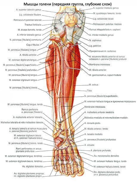
Innervation: deep peroneal nerve(LIV-SI). Blood supply: anterior tibial artery.
Extensor muscle longus toe(m.extensor hallucis longus) is located between the tibialis anterior muscle medially and the extensor digitorum longus muscle laterally; partially covered by them in front. Begins on the middle third of the anterior surface of the fibula, interosseous membrane of the leg. The muscle tendon passes down the dorsum of the foot under the superior and inferior extensor tendon retinaculum in a separate synovial sheath and inserts on the distal phalanx of the big toe. Individual tendon bundles can also attach to the proximal phalanx.
Function: extends the big toe; also participates in the extension of the foot at the ankle joint.
Innervation: deep peroneal nerve (LIV-SI).
Blood supply: anterior tibial artery.
, , ,
Posterior calf muscle group
The muscles of the posterior group form two layers - superficial and deep. The superficial one is more developed triceps lower leg, which creates the characteristic roundness of the lower leg for humans. The deep layer is formed by a small popliteus muscle and 3 long muscles: flexor digitorum longus (located most medially), tibialis posterior (occupies an intermediate position) and flexor hallucis longus (located laterally).

Superficial layer of the posterior muscle group of the lower leg
The triceps surae muscle consists of two muscles - the gastrocnemius muscle, which is located superficially, and the soleus muscle, hidden under the gastrocnemius. Calf muscle refers to the biarticular muscles, it acts on two joints - the knee and ankle, while the soleus muscle is single-joint - it acts only on the ankle joint.
Calf muscle(m.gastrocnemius) has two heads: medial and lateral, the surface layers of which are represented by strong tendon bundles. The lateral head (caput laterale) begins on the outer surface of the lower epiphysis of the femur above the lateral condyle. The medial head (caput mediate) begins on the medial condyle of the femur. Under each head of the gastrocnemius muscle there is a synovial bursa. Between the lateral head and the capsule of the knee joint is located lateral subtendinous bursa of the gastrocnemius muscle(bursa subtendinea musculi gastrocnemii lateralis). Between the medial head and the joint capsule is medial subtendinous bursa of the gastrocnemius muscle(bursa subtendinea musculi gastrocnemii medialis). Both bags, as a rule, communicate with the cavity of the knee joint.
In the middle of the lower leg, both heads of the gastrocnemius muscle pass into a thick tendon, which narrows downwards and merges with the tendon of the soleus muscle, forming the calcaneal (Achilles) tendon (tendo calcaneus, s.Achilli), which is attached to the calcaneal tubercle. Between the tendon and the calcaneus there is a bursa of the calcaneal (Achilles) tendon (bursa tendinis calcanei, s.Achillis).
Soleus muscle(m.soleus) thick, flat, lies under the gastrocnemius muscle. In front of it are the muscles of the deep layer. The soleus muscle has an extensive origin on back surface tibia (on the line of the soleus muscle) and on the tendon arch (arcus tendineus musculi solei), which spreads between the tibia and fibula. The soleus muscle has a feathery structure, passes into a flat tendon, which participates in the formation of the calcaneal tendon.
Function: the triceps muscle flexes the lower leg and foot (plantar flexion); with a fixed foot, it holds the shin on the talus, preventing it from tipping forward.
Innervation: tibial nerve (LIV-SI).
Plantaris muscle
(m.plantaris) is unstable, has a small abdomen and a long thin tendon. It begins on the lateral epicondyle of the femur and on the oblique popliteal ligament. The tendon of this muscle passes between the gastrocnemius and soleus muscles, is adjacent to the medial edge of the calcaneal tendon, together with which it is attached to the calcaneal tubercle.
Function: stretches the capsule of the knee joint, participates in flexion of the lower leg and foot.
Deep layer of the posterior muscle group of the leg
The deep layer is formed by 4 muscles: the popliteus, flexor digitorum longus, flexor hallucis longus and tibialis posterior, which are separated from the soleus muscle by the deep plate of the fascia of the leg.

The popliteal muscle (m.popliteus) lies deep in the popliteal fossa. It begins as a thick tendon on the outer surface of the lateral femoral condyle (below the attachment of the fibular collateral ligament). The muscle is adjacent to the posterior surface of the joint capsule and is located below the arcuate popliteal ligament, where its medial bundles begin. The muscle attaches to a triangular area on the posterior surface of the tibia, above the line of the soleus muscle.
Function: bends the lower leg, turning it inward; stretches the capsule of the knee joint, protecting the synovial membrane from pinching.
Innervation: tibial nerve (LIV-SII).
Blood supply: popliteal artery.

Long flexor digitorum(m.flexor digitorum longus) has a bipinnate structure, begins in fleshy bundles on the posterior surface of the body of the tibia below the line of the soleus muscle, as well as on the fascia and posterior intermuscular septum of the leg. Located behind and medial to the tibialis posterior muscle. The flexor digitorum longus tendon runs downward and crosses the tibialis posterior tendon posteriorly and laterally. The muscle tendon then passes to the sole of the foot behind the medial malleolus under the flexor tendon retinaculum in a separate synovial sheath (between the tibialis posterior tendons medially and the flexor pollicis longus tendon laterally). The tendon then bends around the posterior and inferior support of the talus. Located above the flexor digitorum brevis, it is divided into 4 separate tendons, which are attached to the distal phalanges of the II-V fingers, first piercing the tendons of the flexor digitorum brevis (similar to the tendons of the flexor digitorum profundus on the hand).
Function: bends the distal phalanges of the II-V fingers; bends the foot, turning it outward.
Innervation: tibial nerve (LIV-SII).
Blood supply: posterior tibial artery.
Flexor hallucis longus
(m.flexor hallucus longus) - bipennate muscle, begins on the lower two-thirds of the body of the fibula, interosseous membrane, posterior intermuscular septum of the leg. Located lateral and posterior to the tibialis posterior muscle. The flexor hallucis longus tendon passes under the flexor tendon retinaculum behind the medial malleolus and lateral to the flexor digitorum longus tendon in a separate synovial sheath. Next, the tendon of the long flexor of the big toe lies in the groove of the same name on the posterior process of the talus, passing forward under the support of the talus. Having reached the plantar surface of the big toe, the flexor hallucis longus tendon attaches to its distal phalanx. On its way through the foot, this tendon intersects with (lies underneath) the flexor digitorum longus tendon. Throughout the plantar surface of the first metatarsal bone, the tendon of the flexor hallucis longus lies between the medial and lateral bellies of the flexor hallucis brevis.
Function: flexes the big toe, participates in flexion (supination) and adduction of the foot; strengthens the longitudinal arch of the foot.
Innervation: tibial nerve (LIV-SII).
Blood supply: posterior tibial and peroneal arteries.
The posterior tibialis muscle (m.tibialis posterior) is located deep on the back of the lower leg between the flexor digitorum longus (medially) and the flexor hallucis longus (laterally). Begins on the posterior surface of the body of the fibula (between the medial ridge and the interosseous margin), the lower surface of the lateral condyle and on the upper two-thirds of the body of the tibia (below the line of the soleus muscle) and the interosseous membrane of the tibia.
The muscle continues into a strong tendon, which lies in a groove on the posterior surface of the medial malleolus in front of the flexor digitorum longus tendon (under the retinaculum of the flexor tendons). Moving onto the plantar surface of the foot, the tendon attaches to the tuberosity of the navicular bone, to all 3 wedge-shaped bones, as well as to the base of the IV (sometimes V) metatarsal bone.
Function: flexes the foot (plantar flexion), adducts the foot and supinates it.
Innervation: tibial nerve (LIV-SII).
Blood supply: posterior tibial artery.
Lateral calf muscle group
The lateral group is represented by the long and short peroneal muscles, which are located on the lateral surface of the leg under the fascia between the anterior and posterior intermuscular septa.
The long peroneus muscle (m.peroneus longus) is bipinnate, lies superficially, begins on the head and upper two-thirds of the lateral surface of the fibula, on the lateral condyle of the tibia, the fascia of the leg and on the intermuscular septa of the leg. At the level of the ankle joint, the muscle tendon, bending around the lateral malleolus from behind, passes first under the superior retinaculum of the peroneal tendons in the common synovial sheath with the tendon of the short peroneal muscle, and then in the groove on the calcaneus (under the lower retinaculum of the peroneal tendons). On the sole, the tendon of the peroneus longus muscle passes obliquely forward and medially, lies in the groove of the same name in the cuboid bone in a separate (own) synovial sheath. The tendon is attached to the base of the first and second metatarsal bones and to the medial cuneiform bone.

At those points where the tendon changes direction (behind the lateral malleolus and on the cuboid bone), it usually thickens due to fibrocartilage or sesamoid bone formed in its thickness.
Function: bends the foot, raises its lateral edge (pronation), strengthens the transverse and longitudinal arches of the foot.
Blood supply: lateral inferior genicular artery, peroneal artery.
The short peroneus muscle (m.peroneus brevis) is bipinnate, begins on the lower two-thirds of the lateral surface of the fibula and on the intermuscular septa of the leg. The muscle tendon passes onto the foot behind the lateral malleolus under the retinaculum of the peroneal tendons, lying in the common synovial sheath along with the peroneus longus tendon. At the lower edge of this retinaculum, the peroneus brevis tendon turns forward and passes along the outside of the calcaneus under the fibular trochlea to its insertion on the base of the fifth metatarsal.
Function: raises the lateral edge of the foot; prevents the sole from turning inwards; flexes the foot (plantar flexion).
Innervation: superficial peroneal nerve (LIV-SI).
Blood supply: peroneal artery.
It's full of small muscles like finger extensors and large ones like the soleus muscle.
We will not analyze all the muscles in detail. Let us dwell only on the most basic, most noticeable ones.
Among the muscles of the lower leg, there are anterior, lateral and posterior muscle groups. The anterior group includes mainly extensors of the foot, the lateral group includes flexors and foot muscles, and the posterior group includes flexors and supina.Calf muscles front view :
1 - peroneus longus muscle;
2 - medial head of the gastrocnemius muscle;
3 - tibialis anterior muscle;
4 - soleus muscle;
5 - short peroneus muscle;
6 - extensor digitorum longus;
7 - superior extensor retinaculum;
8 - tendon of the anterior tibialis muscle;
9 - lower extensor retinaculum
Front group
(m. tibialis anterior) extends and adducts the foot, raising its medial edge. A long, narrow, superficial muscle whose origin is located on the lateral condyle of the tibia and the interosseous membrane.
The attachment site is located on the plantar surface of the medial sphenoid bone and on the base of the first metatarsal bone. The subtendinous bursa of the tibialis anterior muscle is also located here (bursa subtendinea m. tibialis anterioris).
The long extensor digitorum (m. extensor digitorum longus) extends the II–V fingers, as well as the foot, lifting its lateral (outer) edge together with the third peroneal muscle. The muscle begins from the upper epiphysis of the tibia, the head and anterior edge of the fibula and the interosseous membrane. The muscle passes into a long, narrow tendon, which divides into five thin individual tendons. Four of them are attached to the back of the II–IV fingers in such a way that the middle bundles of tendons are attached to the base of the middle phalanx, and the lateral bundles to the base of the distal phalanx. The fifth tendon attaches to the base of the fifth metatarsal bone.

1 - articular muscle of the knee;
2 - quadratus femoris muscle;
3 - short peroneus muscle;
4 - long extensor of the big toe;
5 - short extensor of the big toe;
6 - tendon of the long extensor of the big toe;
7 - extensor digitorum brevis
The long extensor hallucis longus (m. extensor hallucis longus) extends the big toe, as well as the foot itself, raising its medial edge. Partially covered by the two previous muscles, located between them. Its point of origin is the lower part of the medial surface of the body of the fibula, and the point of attachment is the base of the distal phalanx. Part of the tendon bundles fuses with the base of the proximal phalanx.
Lateral group
The long peroneus muscle (m. peroneus longus) abducts and flexes the foot, lowering its medial edge. Located on the lateral surface of the lower leg. The muscle begins from the head and upper part of the body of the fibula and is attached to the medial sphenoid bone and the base of the I–II metatarsal bones.

Calf muscles (back view):
1 - plantaris muscle;
2 - gastrocnemius muscle: a) medial head, b) lateral head;
3 - soleus muscle;
4 - fascia of the leg;
5 - tendon of the posterior tibial muscle;
7 - flexor digitorum longus tendon;
8 - calcaneal tendon (Achilles tendon)
Back group
Back group includes two muscle groups.
Surface layer
Triceps surae muscle(m. triceps surae) bends the lower leg at the knee joint, bends and rotates the foot outward. When the foot is in a fixed position, the lower leg and thigh are pulled posteriorly. The muscle consists of the superficial gastrocnemius muscle and the deep soleus muscle. (m. gastrocnemius) has two heads. The medial head (caput mediale) starts from the medial epicondyle of the femur, and the lateral head (caput laterale) starts from the lateral epicondyle. Both heads are connected into a common tendon and attached to the calcaneal tubercle.
(m. soleus) is covered by the gastrocnemius muscle, starts from the head and upper third of the posterior surface of the body of the fibula and from the line of the soleus muscle of the tibia. The muscle is attached to the calcaneal tubercle, fused with the tendon of the gastrocnemius muscle. The common tendon in the lower third of the leg forms the calcaneal tendon (tendo calcaneus), the so-called Achilles tendon. The mucous bursa of the heel tendon (bursa tendinis calcanei) is also located here.
Plantaris muscle(m. plantaris) stretches the capsule of the knee joint when flexing and rotating the tibia. The muscle is rudimentary and unstable, has a spindle-shaped shape. Its point of origin is located on the lateral condyle of the femur and the bursa of the knee joint, and its attachment point is on the calcaneus.
|
|
Calf muscles (back view): 1 - plantaris muscle; 2 - popliteus muscle; 3 - soleus muscle; 4 - tendon of the plantaris muscle; 5 - gastrocnemius muscle: a) medial head, b) lateral head; 6 - tendon of the long peroneus muscle; 7 - tendon of the posterior tibial muscle; 8 - short peroneus muscle; 9 - flexor digitorum longus tendon; 10 - calcaneal tendon (Achilles tendon) |
|
|
Calf muscles (back view): 1 - popliteus muscle; 2 - soleus muscle; 4 - peroneus longus muscle; 5 - flexor digitorum longus; 6 - flexor pollicis longus; 7 - short peroneus muscle; 8 - flexor retinaculum; 9 - superior retinaculum of the peroneus longus and brevis muscles |


1 - popliteus muscle;
2 - short peroneus muscle;
3 - tibialis posterior muscle;
4 - short flexor of the big toe;
5 - short flexor of the little toe;
6 - flexor digitorum longus tendon;
7 - interosseous muscles
Deep layer
Hamstring muscle(m. popliteus) bends the lower leg, rotating it inward and pulling the capsule of the knee joint. A short flat muscle, located on the posterior surface of the knee joint capsule, starts from it and from the lateral condyle of the femur, and is attached to the posterior surface of the body of the tibia.
Flexor digitorum longus(m. flexor digitorum longus) bends the distal phalanges of the II–V fingers and takes part in the outward rotation of the foot, raising its medial edge. It is located on the posterior surface of the tibia, starting from the middle third of the posterior surface of the body of the tibia and from the deep sheet of the fascia of the leg. The muscle tendon is divided into four tendons, which are attached to the base of the distal phalanges of the II–V fingers.
Flexor pollicis longus(m. flexor hallucis longus) flexes the big toe, takes part in the flexion of fingers II–V thanks to fibrous bundles, which are a continuation of the tendon, and also flexes and rotates the foot.
The muscle starts from the lower two-thirds of the posterior surface of the body of the fibula and from the interosseous membrane, and is attached to the base of the distal phalanx of the thumb.
(m. tibialis posterior) flexes and adducts the foot, rotating it outward. It is located on the interosseous membrane between the two previous muscles and is partially covered by the flexor pollicis longus. Its point of origin is on the posterior surfaces of the bodies of the tibia and fibula, and its attachment point is on the wedge-shaped bones of the foot and the tuberosity of the navicular bone.
Posterior muscle group of the lower leg.
Superficial layer (calf muscles):
M. triceps surae, triceps surae muscle, forms the main mass of the calf elevation. It consists of two muscles - m. gastrocnemius, located superficially, and m. soleus, lying under it; both muscles below have one common tendon.
- M. gastrocnemius, the gastrocnemius muscle, starts from the facies poplitea of the femur behind both condyles with two heads, which, with their tendon origin, fuse with the capsule of the knee joint. The heads pass into the tendon, which, merging with the tendon m. soleus, continues into the massive Achilles tendon, tendo calcaneus (Achillis), attached to the posterior surface of the tubercle of the calcaneus. At the point of attachment between the tendon and the bone there is a very permanent synovial bursa, bursa tendinis calcanei (Achillis).
- M. soleus, soleus muscle, thick and fleshy. It lies under the calf muscle, occupying a large area on the bones of the lower leg. The line of its origin is located on the head and on the upper third of the posterior surface of the fibula and descends along the tibia almost to the border of the middle third of the tibia with the lower one. In the place where the muscle spreads from the fibula to the tibia, a tendon arch is formed, arcus tendineus m. solei, under which the popliteal artery and n. tibialis. Tendon sprain m. soleus merges with the Achilles tendon.

M. plantaris, plantaris muscle. It originates from the facies poplitea above the lateral condyle of the femur and from the capsule of the knee joint, soon passing into a very long and thin tendon that stretches in front of the m. gastrocnemius and attaches to the calcaneal tubercle. This muscle undergoes reduction and in humans is a rudimentary formation, as a result of which it may be absent. Function. All muscles m. triceps surae (including m. plantaris) produces flexion at the ankle joint both with the free leg and with support on the end of the foot. Since the line of pull of the muscle passes medially to the axis of the subtalar joint, it also causes adduction of the foot and supination. When standing, the triceps surae (especially the m. soleus) prevents the body from tipping forward at the ankle joint. The muscle has to work primarily when burdened by the weight of the whole body, and therefore it is strong and has a large physiological diameter; m. gastrocnemius, as a biarticular muscle, can also flex the knee when the lower leg and foot are strengthened. (Inn. m. triceps surae and m. plantaris - L5-S2. N. tibialis.) The deep layer, separated from the superficial by the deep fascia of the leg, is composed of three flexors, which oppose the three homonymous extensors lying on the anterior surface of the leg.

M. flexor digitorum longus, long flexor of the fingers, the most medial of the muscles of the deep layer. It lies on the posterior surface of the tibia, from which it originates. The tendon of the muscle descends behind the medial malleolus, in the middle of the sole it divides into four secondary tendons, which go to the four fingers II-V, pierce the tendon m. flexor digitorum brevis and are attached to the distal phalanges. The function in terms of bending the fingers is small; the muscle mainly acts on the foot as a whole, producing flexion and supination when the leg is free. She also, together with m. triceps surae is involved in placing the foot on the toe (walking on tiptoes). When standing, the muscle actively helps strengthen the arch of the foot in the longitudinal direction. When walking, presses fingers to the ground. (Inn. L5-S1. N. tibialis.)

M. tibialis posterior, tibialis posterior muscle, occupies the space between the bones of the leg, lying on the interosseous membrane and partly on the tibia and fibula. From these places the muscle receives its initial fibers, then with its tendon it bends around the medial malleolus and, reaching the sole, is attached to the tuberositas ossis navicularis, and then by several bundles to the three wedge-shaped bones and the bases of the II-IV metatarsal bones. Function. Bends the foot and brings it together with m. tibialis anterior. Together with other muscles also attached to the medial edge of the foot (m. tibialis anterior et m. peroneus longus), m. tibialis posterior forms a kind of stirrup, which strengthens the arch of the foot; stretching its tendon through the lig. calcaneonavicular, the muscle supports the head of the talus together with this ligament. (Inn. L5-S1. N. tibialis.)
M. flexor hallucis longus, long flexor of the big toe, the most lateral of the deep layer muscles. Lies on the posterior surface of the fibula, from which it originates; the tendon runs in a groove on the processus posterior of the talus, approaches the sustentaculum tali to the big toe, where it attaches to its distal phalanx. Function. Flexes the thumb, and also due to a possible connection with the tendon of the m. flexor digitorum longus can act in the same sense on Pi even on fingers III and IV. Like the rest of the posterior muscles of the leg, m. flexor hallucis longus produces flexion, adduction and supination of the foot and strengthens the arch of the foot in the anteroposterior! direction. (Inn. L5-S2. N. tibialis.)
Superficial layer (calf muscles):
1. M. triceps surae, triceps surae muscle, forms the main mass of the calf elevation. It consists of two muscles - m. gastrocnemius, located superficially, and m. soleus, lying under it; both muscles below have one common tendon.
M. gastrocnemius, gastrocnemius muscle, starts from the facies poplitea of the femur behind both condyles with two heads, which, with their tendon origin, fuse with the capsule of the knee joint. The heads pass into the tendon, which, merging with the tendon m. soleus, continues into the massive Achilles tendon, tendo calcaneus (Achillis), attached to the posterior surface of the tubercle of the calcaneus.
At the point of attachment between the tendon and the bone there is a very permanent synovial bursa, bursa tendinis calcanei (Achillis).
M. soleus, soleus muscle, thick and meaty. It lies under the calf muscle, occupying a large area on the bones of the lower leg. The line of its origin is located on the head and on the upper third of the posterior surface of the fibula and descends along the tibia almost to the border of the middle third of the tibia with the lower one. In the place where the muscle spreads from the fibula to the tibia, a tendon arch is formed, arcus tendineus m. solei, under which the popliteal artery and n. tibialis. Tendon sprain m. soleus merges with the Achilles tendon.
2. M. plantaris, plantaris muscle. It originates from the facies poplitea above the lateral condyle of the femur and from the capsule of the knee joint, soon passing into a very long and thin tendon that stretches in front of the m. gastrocnemius and attaches to the calcaneal tubercle. This muscle undergoes reduction and in humans is a rudimentary formation, as a result of which it may be absent.
Function. All muscles m. triceps surae (including m. plantaris) produces flexion at the ankle joint both with the free leg and with support on the end of the foot. Since the line of pull of the muscle passes medially to the axis of the subtalar joint, it also causes adduction of the foot and supination. When standing, the triceps surae (especially the m. soleus) prevents the body from tipping forward at the ankle joint.
The muscle has to work primarily when burdened by the weight of the whole body, and therefore it is strong and has a large physiological diameter; m. gastrocnemius, as a biarticular muscle, can also flex the knee when the lower leg and foot are strengthened. (Inn. m. triceps surae and m. plantaris - LV - SII. N. tibialis.)
Deep layer, separated from the superficial by the deep fascia of the leg, is composed of three flexors, which oppose the three extensors lying on the anterior surface of the leg.

3. M. flexor digitorum longus, long flexor of the fingers, the most medial of the muscles of the deep layer. It lies on the posterior surface of the tibia, from which it originates. The tendon of the muscle descends behind the medial malleolus, in the middle of the sole it divides into four secondary tendons, which go to the four fingers I-V, perforate like the deep flexor on the hand tendon m. flexor digitorum brevis and are attached to the distal phalanges.
Function in terms of finger flexion, it is small; the muscle mainly acts on the foot as a whole, producing flexion and supination when the leg is free. She also, together with m. triceps surae is involved in placing the foot on the toe (walking on tiptoes). When standing, the muscle actively helps strengthen the arch of the foot in the longitudinal direction. When walking, presses fingers to the ground. (Inn. LV - SI. N. tibialis.)
4. M. tibialis posterior, posterior tibialis muscle, occupies the space between the bones of the leg, lying on the interosseous membrane and partly on the tibia and fibula. From these places the muscle receives its initial fibers, then with its tendon it bends around the medial malleolus and, reaching the sole, is attached to the tuberositas ossis navicularis, and then by several bundles to the three wedge-shaped bones and the bases of the II-IV metatarsal bones.
Function. Bends the foot and brings it together with m. tibialis anterior. Together with other muscles also attached to the medial edge of the foot (m. tibialis anterior et m. peroneus longus), m. tibialis posterior forms a kind of stirrup, which strengthens the arch of the foot; stretching its tendon through the lig. calcaneonavicular, the muscle supports the head of the talus together with this ligament. (Inn. LV - SI. N. tibialis.)

5. M. flexor hallucis longus, long flexor of the big toe, the most lateral of the deep layer muscles. Lies on the posterior surface of the fibula, from which it originates; the tendon runs in a groove on the processus posterior of the talus, approaches under the sustentaculum tali to the big toe, where it attaches to its distal phalanx.
Function. Flexes the thumb, and also due to a possible connection with the tendon of the m. flexor digitorum longus can act in the same sense on fingers II and even III and IV. Like the rest of the posterior muscles of the leg, m. flexor hallucis longus produces flexion, adduction and supination of the foot and strengthens the arch of the foot in the anteroposterior! direction. (Inn. N. tibialis.)

Shin refers to lower limb. It is located between the foot and the knee area. The lower leg is formed by two bones - the fibula and the tibia. They are surrounded by muscle fibers on three sides. The muscles of the lower leg, the anatomy of which will be discussed later, move the fingers and foot.
Tibia
This element has an extension at the top edge. In this area, condyles are formed: lateral and medial. On top of them are the surfaces of the joints. They articulate with the femoral condyles. On the outside of the lateral segment there is an articular surface through which the connection occurs with the head of the fibula. The body of the tibial element looks like a triangular prism. Its base is directed posteriorly and has, respectively, 3 surfaces: posterior, external and internal. Between the last two there is an edge. It's called the front one. In its upper part it passes into the tibial tuberosity. This area is intended for fixation. The lower part of the tibia has an extension, and on inner surface there is a protrusion. It is oriented downward. This projection is called the medial malleolus. On the back side of the bone lies a rough segment of the soleus muscle. The distal epiphysis contains the articular surface. It serves to connect with

Second element
The fibula is thin, long, and located laterally. Its upper end has a thickening - a head. It connects to the tibia. Lower section the element is also thickened and forms the lateral malleolus. It, like the head of the fibula, is oriented outward and can be easily palpated.
Calf muscles: their location, functions
The fibers are located on three sides. Highlight different muscles shins. The anterior group performs extension of the foot and toes, supination and adduction of the foot. This segment includes three types of fibers. The tibialis anterior muscle of the leg is the first to be formed. The remaining fibers form the long extensors of the fingers and a separate one for the big toe. The posterior group of muscles of the lower leg forms a larger number of fibers. In particular, there are long flexors of the fingers and, separately, for the thumb, popliteus, and triceps surae muscles. Tibial fibers also lie here. TO outdoor group include the short and long peroneus muscles of the leg. These fibers flex, pronate and abduct the foot.
Tibial segment
This anterior muscle of the leg begins from the bone of the same name, its outer surface, fascia and interosseous membrane. They are directed downwards. The fibers pass under two ligaments. They are located in the ankle area. These areas - the upper and lower retinaculum of the extensor tendons - are represented by places of thickening of the fascia of the foot and lower leg. The site of attachment of the fibers is the medial wedge and the base of the metatarsal (first) bone. The muscle can be palpated quite well along its entire length, especially in the area where it transitions to the foot. In this place, its tendon protrudes during extension. The task of this calf muscle is to supinate the foot.

Extensor digitorum (long)
It runs from the anterior muscle outward into upper area shins. Its fibers begin from the head and marginal areas of the tibia, fascia and interosseous membrane. The extensor, moving to the foot, is divided into five tendons. Four are attached to the distal ones (second to fifth), the last one to the base of the 5th metatarsal. The task of the extensor, acting as a multi-joint muscle of the leg, is not only to coordinate the extension of the fingers, but also the foot. Due to the fact that one tendon is fixed at its edge, the fibers also pronate the area somewhat.
Extensors of the thumbs
The fibers begin in the area of the lower leg from the interosseous membrane and the inner part of the fibula. The extensors have less strength than the segments described above. The site of attachment of this is the distal phalanges in thumbs. These muscles of the lower leg not only extend them, but also the feet, also contributing to their supination.

Flexor digitorum (long)
It starts from the back of the tibia, passing under the medial malleolus onto the foot. The channel for it is located under the retinaculum. Next, the muscle is divided into four segments. On the foot (its plantar surface), fibers cross the tendon from the flexor (long) hallux. Then they are joined by the quadratus plantae muscle. Four formed tendons are fixed to the distal phalanges (at their base) of 2-5 fingers. The task of this muscle is, among other things, to flex and supinate the foot. The fibers of the quadrate segment are attached to the tendon. Due to this, the muscle action is averaged. Lying under the medial malleolus and dividing fan-shaped towards the phalanges, the long flexor also provokes some adduction of the fingers to the median surface of the body. By pulling quadratus muscle tendons, this effect is slightly reduced.
Triceps surae muscle
It runs along the back surface and has 3 heads. Two form the superficial section - the gastrocnemius muscle, from the third - the deep one - the fibers of the soleus segment depart. All heads connect and form the common Achilles (heel) tendon. It is attached to the tubercle of the corresponding bone. The gastrocnemius muscle starts from the femoral condyles: lateral and medial. The purpose of the two heads located in this area is twofold. They coordinate flexion at the knee joint and foot flexion at the ankle. The medial element descends slightly lower and is better developed than the lateral one. The soleus muscle extends from the posterior side of the upper third of the tibia. It is also attached to the arch of tendon located between the bones. The fibers run somewhat lower and deeper than the calf. They lie behind the subtalar and cause flexion of the foot. The triceps muscle can be felt under the skin. The calcaneal tendon protrudes posteriorly from the transverse axis of the ankle joint. Due to this, the triceps muscle has a large torque relative to this line. The heads of the gastrocnemius segment participate in the formation of the rhomboid popliteal fossa. Its boundaries are: two-headed femoral muscle(outside and above), semimembranous fibers (inside and above), plantar and two heads of the gastrocnemius segment (below). The bottom of the fossa is formed by the capsule of the knee joint and the vessels and nerves that supply the foot and lower leg run through this area.

Flexor pollicis longus
This muscle of the posterior surface of the leg is characterized greatest strength. On the plantar side of the foot, fibers run between the heads from a short segment responsible for flexion of the big toe. The muscle begins from the posterior side (lower part) of the fibula and the intermuscular septum (posterior). The site of fixation is the plantar surface of the base of the distal phalanx in the big toe. Due to the fact that the tendon of the muscle partially passes into the long flexor element of the same name, it has some influence on the movements of 2-3 fingers. The presence of 2 large sesamoid bone elements on the surface of the sole of the metatarsophalangeal joint provides an increase in the moment of rotation of the fibers. The tasks of the segment include flexion of the entire foot and big toe.
Second division of tibial fibers
This posterior segment is located under the triceps muscle. The fibers begin from the interosseous membrane and the areas of the fibula and tibia adjacent to it. The site of attachment of the muscle is the tubercle of the scaphoid, the base of the metatarsals and all wedge-shaped elements. The muscle lies under the medial malleolus and performs flexion of the foot, supination and adduction. A canal passes between the soleus and tibial fibers. It is presented in the form of a slit. Nerves and blood vessels pass through it.

Popliteal segment
It is formed flat short fibers. The muscle is directly adjacent to knee joint behind. The fibers originate from the femoral condyle (lateral), below the gastrocnemius, and the bursa of the knee joint. They pass down and attach above the soleus muscle to the tibia. Because the fibers are partially attached to the joint capsule, they pull it posteriorly when flexed. The task of the muscle is to pronate and flex the lower leg.
Long fibular segment
This muscle has a feathery structure. It runs along the surface of the fibula. It starts from its head, the condyle of the tibial element, partly from the fascia. It is also attached to the 2-thirds area outside fibula. When the muscle contracts, abduction, pronation and flexion of the foot occur. The tendon of the long peroneal segment passes posteriorly and inferiorly around the lateral malleolus. In the area of the heel bone there are ligaments - the upper and lower retinaculum. When moving to the plantar part of the foot, the tendon runs along the groove. It is located on the underside of the cuboid bone. The muscle reaches the inside of the foot.

Short fibular fibers
The tendon of the segment bends around the lateral malleolus posteriorly and inferiorly. It is attached to the tubercle on the 5th metatarsal bone. The segment begins from the intermuscular septa and the outer part of the fibula. The task of the fibers is abduction, pronation and flexion of the foot.


